 Movie
Movie Controller

[English] 日本語
 Yorodumi
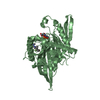
- PDB-3b8k: Structure of the Truncated Human Dihydrolipoyl Acetyltransferase (E2) -
+ Open data

- Basic information
| Entry | Database: PDB / ID: 3b8k | ||||||
|---|---|---|---|---|---|---|---|
| Title | Structure of the Truncated Human Dihydrolipoyl Acetyltransferase (E2) | ||||||
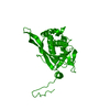
 Components Components | Dihydrolipoyllysine-residue acetyltransferase Dihydrolipoyl transacetylase Dihydrolipoyl transacetylase | ||||||
 Keywords Keywords | TRANSFERASE / central beta-sheet surrounded by five alpha-helices | ||||||
| Function / homology |  Function and homology informationdihydrolipoyllysine-residue acetyltransferase / dihydrolipoyllysine-residue acetyltransferase activity / acetyl-CoA biosynthetic process from pyruvate / pyruvate dehydrogenase complex / : / Pyruvate metabolism / Glyoxylate metabolism and glycine degradation / Regulation of pyruvate dehydrogenase (PDH) complex / Signaling by Retinoic Acid / tricarboxylic acid cycle ...dihydrolipoyllysine-residue acetyltransferase / dihydrolipoyllysine-residue acetyltransferase activity / acetyl-CoA biosynthetic process from pyruvate / pyruvate dehydrogenase complex / : / Pyruvate metabolism / Glyoxylate metabolism and glycine degradation / Regulation of pyruvate dehydrogenase (PDH) complex / Signaling by Retinoic Acid / tricarboxylic acid cycle / glucose metabolic process / mitochondrial matrix / intracellular membrane-bounded organelle / mitochondrion / identical protein binding Function and homology informationdihydrolipoyllysine-residue acetyltransferase / dihydrolipoyllysine-residue acetyltransferase activity / acetyl-CoA biosynthetic process from pyruvate / pyruvate dehydrogenase complex / : / Pyruvate metabolism / Glyoxylate metabolism and glycine degradation / Regulation of pyruvate dehydrogenase (PDH) complex / Signaling by Retinoic Acid / tricarboxylic acid cycle ...dihydrolipoyllysine-residue acetyltransferase / dihydrolipoyllysine-residue acetyltransferase activity / acetyl-CoA biosynthetic process from pyruvate / pyruvate dehydrogenase complex / : / Pyruvate metabolism / Glyoxylate metabolism and glycine degradation / Regulation of pyruvate dehydrogenase (PDH) complex / Signaling by Retinoic Acid / tricarboxylic acid cycle / glucose metabolic process / mitochondrial matrix / intracellular membrane-bounded organelle / mitochondrion / identical protein bindingSimilarity search - Function | ||||||
| Biological species |  Homo sapiens (human) Homo sapiens (human) | ||||||
| Method | ELECTRON MICROSCOPY / single particle reconstruction / cryo EM / Resolution: 8.8 Å | ||||||
 Authors Authors | Yu, X. / Hiromasa, Y. / Tsen, H. / Stoops, J.K. / Roche, T.E. / Zhou, Z.H. | ||||||
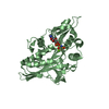
 Citation Citation | Journal: Structure / Year: 2008 Title: Structures of the human pyruvate dehydrogenase complex cores: a highly conserved catalytic center with flexible N-terminal domains. Authors: Xuekui Yu / Yasuaki Hiromasa / Hua Tsen / James K Stoops / Thomas E Roche / Z Hong Zhou /  Abstract: Dihydrolipoyl acetyltransferase (E2) is the central component of pyruvate dehydrogenase complex (PDC), which converts pyruvate to acetyl-CoA. Structural comparison by cryo-electron microscopy (cryo- ...Dihydrolipoyl acetyltransferase (E2) is the central component of pyruvate dehydrogenase complex (PDC), which converts pyruvate to acetyl-CoA. Structural comparison by cryo-electron microscopy (cryo-EM) of the human full-length and truncated E2 (tE2) cores revealed flexible linkers emanating from the edges of trimers of the internal catalytic domains. Using the secondary structure constraints revealed in our 8 A cryo-EM reconstruction and the prokaryotic tE2 atomic structure as a template, we derived a pseudo atomic model of human tE2. The active sites are conserved between prokaryotic tE2 and human tE2. However, marked structural differences are apparent in the hairpin domain and in the N-terminal helix connected to the flexible linker. These permutations away from the catalytic center likely impart structures needed to integrate a second component into the inner core and provide a sturdy base for the linker that holds the pyruvate dehydrogenase for access by the E2-bound regulatory kinase/phosphatase components in humans. | ||||||
| History |
|
- Structure visualization
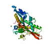
| Movie |
Movie viewer |
|---|---|
| Structure viewer | Molecule: MolmilJmol/JSmol |
- Downloads & links
-Download
| PDBx/mmCIF format | 3b8k.cif.gz | 52.5 KB | Display | PDBx/mmCIF format |
|---|---|---|---|---|
| PDB format | pdb3b8k.ent.gz | 36.7 KB | Display | PDB format |
| PDBx/mmJSON format | 3b8k.json.gz | Tree view | PDBx/mmJSON format | |
| Others |  Other downloads Other downloads |
-Validation report
| Arichive directory | https://data.pdbj.org/pub/pdb/validation_reports/b8/3b8kftp://data.pdbj.org/pub/pdb/validation_reports/b8/3b8k | HTTPS FTP |
|---|
-Related structure data
| Related structure data | 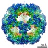 1448MC M: map data used to model this data C: citing same article ( |
|---|---|
| Similar structure data |


-Links
 PDBj
PDBj


- Assembly
| Deposited unit | 
|
|---|---|
| 1 |
|
-Components
| #1: Protein | Dihydrolipoyl transacetylase / E.C.2.3.1.12 / Pyruvate dehydrogenase complex E2 subunit / PDCE2 / E2 / Dihydrolipoamide S- acetyltransferase ...Pyruvate dehydrogenase complex E2 subunit / PDCE2 / E2 / Dihydrolipoamide S- acetyltransferase component of pyruvate dehydrogenase complex / PDC-E2 / 70 kDa mitochondrial autoantigen of primary biliary cirrhosis / PBC / M2 antigen complex 70 kDa subunit Mass: 25938.164 Da / Num. of mol.: 1 / Fragment: C-TERMINAL CATALYTIC DOMAIN Source method: isolated from a genetically manipulated source Details: component of pyruvate dehydrogenase complex, mitochondrial Source: (gene. exp.) Homo sapiens (human) / Gene: DLAT, DLTA / Species (production host): Escherichia coli / Production host:  Escherichia coli BL21(DE3) (bacteria) / Strain (production host): BL21(DE3) Escherichia coli BL21(DE3) (bacteria) / Strain (production host): BL21(DE3)References: UniProt: P10515, dihydrolipoyllysine-residue acetyltransferase |
|---|
-Experimental details
-Experiment
| Experiment | Method: ELECTRON MICROSCOPY |
|---|---|
| EM experiment | Aggregation state: PARTICLE / 3D reconstruction method: single particle reconstruction |
- Sample preparation
| Component | Name: human tE2 / Type: COMPLEX Details: Dodecahedron. Human tE2 was prepared from scE2, which contains a PreScission site in the third linker region. Treatment of scE2 with the PreScission protease (Amersham Biosciences) removed ...Details: Dodecahedron. Human tE2 was prepared from scE2, which contains a PreScission site in the third linker region. Treatment of scE2 with the PreScission protease (Amersham Biosciences) removed the N-terminal 319 amino acids. The resulting tE2 was purified by gel filtration with Sephacryl S-300HR. The assembly of the recombinant molecules into fully functional, pentagonal dodecahedral cores was confirmed by analytical ultracentrifugation |
|---|---|
| Buffer solution | Name: PBS / pH: 7.2 / Details: PBS |
| Specimen | Conc.: 0.2 mg/ml / Embedding applied: NO / Shadowing applied: NO / Staining applied: NO / Vitrification applied: YES |
| Specimen support | Details: This grid plus sample was kept at -170 deg C during imaging |
| Vitrification | Instrument: HOMEMADE PLUNGER / Cryogen name: ETHANE / Details: flash freezing in liquid ethane |
- Electron microscopy imaging
| Microscopy | Model: JEOL 2010F / Date: Oct 1, 2003 |
|---|---|
| Electron gun | Electron source: FIELD EMISSION GUN / Accelerating voltage: 200 kV / Illumination mode: FLOOD BEAM |
| Electron lens | Mode: BRIGHT FIELDBright-field microscopy / Nominal magnification: 69250 X / Calibrated magnification: 69250 X / Nominal defocus max: 2100 nm / Nominal defocus min: 600 nm / Cs: 1 mm |
| Specimen holder | Temperature: 100 K |
| Image recording | Electron dose: 12 e/Å2 / Film or detector model: GATAN ULTRASCAN 4000 (4k x 4k) / Details: 4kx4k |
- Processing
| EM software |
| ||||||||||||
|---|---|---|---|---|---|---|---|---|---|---|---|---|---|
| CTF correction | Details: CTF correction of each image | ||||||||||||
| Symmetry | Point symmetry: I (icosahedral) | ||||||||||||
| 3D reconstruction | Method: cross-common lines / Resolution: 8.8 Å / Num. of particles: 2432 / Nominal pixel size: 2.17 Å / Actual pixel size: 2.17 Å Details: Orientation determination and 3D reconstruction were performed using the IMIRS software package on multiprocessor MS Windows XP computer workstations. The orientations were first estimated ...Details: Orientation determination and 3D reconstruction were performed using the IMIRS software package on multiprocessor MS Windows XP computer workstations. The orientations were first estimated from the particle images in the far-from-focus micrographs and refined to about 30- resolution. These orientation parameters were then further refined using the particles in the close-to-focus micrographs. Symmetry type: POINT | ||||||||||||
| Atomic model building | B value: 30 / Protocol: RIGID BODY FIT / Space: REAL / Target criteria: best fit using the program CHIMERA / Details: REFINEMENT PROTOCOL--rigid body | ||||||||||||
| Atomic model building | PDB-ID: 1EAA Accession code: 1EAA / Details: Homology model based on PBD ID 1eaa / Source name: PDB / Type: experimental model | ||||||||||||
| Refinement step | Cycle: LAST
|
