 Movie
Movie Controller


+ Open data

- Basic information
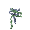
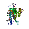
| Entry | Database: PDB / ID: 3b62 | ||||||
|---|---|---|---|---|---|---|---|
| Title | EmrE multidrug transporter in complex with P4P, P21 crystal form | ||||||
 Components Components | Multidrug transporter emrE | ||||||
 Keywords Keywords |  MEMBRANE PROTEIN / HELICAL MEMBRANE PROTEIN / MULTIDRUG RESISTANCE TRANSPORTER / SMR / Antiport / Inner membrane / Transmembrane MEMBRANE PROTEIN / HELICAL MEMBRANE PROTEIN / MULTIDRUG RESISTANCE TRANSPORTER / SMR / Antiport / Inner membrane / Transmembrane | ||||||
| Function / homology |  Function and homology information Function and homology informationEmrE multidrug transporter complex / amino-acid betaine transmembrane transporter activity / choline transmembrane transporter activity / glycine betaine transport / choline transport / xenobiotic detoxification by transmembrane export across the plasma membrane / xenobiotic transport / antiporter activity / response to osmotic stress / xenobiotic transmembrane transporter activity ...EmrE multidrug transporter complex / amino-acid betaine transmembrane transporter activity / choline transmembrane transporter activity / glycine betaine transport / choline transport / xenobiotic detoxification by transmembrane export across the plasma membrane / xenobiotic transport / antiporter activity / response to osmotic stress / xenobiotic transmembrane transporter activity / transmembrane transporter activity / xenobiotic metabolic process / transmembrane transport / cellular response to xenobiotic stimulus / response to xenobiotic stimulus / DNA damage response / membrane / identical protein binding / plasma membraneSimilarity search - Function | ||||||
| Biological species |  Escherichia coli K12 (bacteria) Escherichia coli K12 (bacteria) | ||||||
| Method | X-RAY DIFFRACTION / SYNCHROTRON / MAD / Resolution: 4.4 Å | ||||||
 Authors Authors | Chang, G. / Chen, Y.J. | ||||||
 Citation Citation | Journal: Proc.Natl.Acad.Sci.Usa / Year: 2007 Title: X-ray structure of EmrE supports dual topology model. Authors: Chen, Y.J. / Pornillos, O. / Lieu, S. / Ma, C. / Chen, A.P. / Chang, G. | ||||||
| History |
|
- Structure visualization
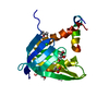
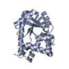
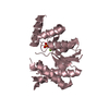
| Structure viewer | Molecule: MolmilJmol/JSmol |
|---|
- Downloads & links
-Download
| PDBx/mmCIF format | 3b62.cif.gz | 24.8 KB | Display | PDBx/mmCIF format |
|---|---|---|---|---|
| PDB format | pdb3b62.ent.gz | 13.6 KB | Display | PDB format |
| PDBx/mmJSON format | 3b62.json.gz | Tree view | PDBx/mmJSON format | |
| Others |  Other downloads Other downloads |
-Validation report
| Arichive directory | https://data.pdbj.org/pub/pdb/validation_reports/b6/3b62ftp://data.pdbj.org/pub/pdb/validation_reports/b6/3b62 | HTTPS FTP |
|---|
-Related structure data


-Links
 PDBj
PDBj
- Assembly
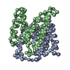
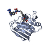
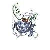
| Deposited unit | 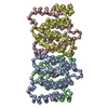
| ||||||||
|---|---|---|---|---|---|---|---|---|---|
| 1 | 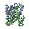
| ||||||||
| 2 | 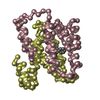
| ||||||||
| Unit cell |
|
-Components
| #1: Protein | Mass: 11963.278 Da / Num. of mol.: 4 Source method: isolated from a genetically manipulated source Source: (gene. exp.) Escherichia coli K12 (bacteria) / Species: Escherichia coli / Strain: K-12 / Gene: emrE, eb, mvrC / Plasmid: pIVEX / Species (production host): Escherichia coli / Production host: Escherichia coli BL21(DE3) (bacteria) / Strain (production host): BL21(DE3) / References: UniProt: P23895#2: Chemical | Tetraphenylphosphonium chloride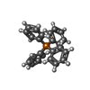  Mass: 339.389 Da / Num. of mol.: 2 / Source method: obtained synthetically / Formula: C24H20P Mass: 339.389 Da / Num. of mol.: 2 / Source method: obtained synthetically / Formula: C24H20P |
|---|
-Experimental details
-Experiment
| Experiment | Method: X-RAY DIFFRACTION / Number of used crystals: 1 |
|---|
- Sample preparation
| Crystal | Density Matthews: 3.72 Å3/Da / Density % sol: 66.95 % |
|---|---|
| Crystal grow | Temperature: 298 K / Method: vapor diffusion, sitting drop / pH: 6.8 Details: 100-200 mM calcium chloride, 100 mM Tris, 11-14% (w/v) PEG 2,000 MME, and 0.3-0.6% (w/v) NG, pH 6.8, VAPOR DIFFUSION, SITTING DROP, temperature 298K |
-Data collection
| Diffraction | Mean temperature: 100 K | ||||||||||||
|---|---|---|---|---|---|---|---|---|---|---|---|---|---|
| Diffraction source | Source: SYNCHROTRON / Site: SSRL  / Beamline: BL11-1 / Wavelength: 0.9790, 0.9793, 0.9184 / Beamline: BL11-1 / Wavelength: 0.9790, 0.9793, 0.9184 | ||||||||||||
| Detector | Type: ADSC QUANTUM 315 / Detector: CCD / Date: Dec 21, 2006 | ||||||||||||
| Radiation | Protocol: MAD / Monochromatic (M) / Laue (L): M / Scattering type: x-ray | ||||||||||||
| Radiation wavelength |
| ||||||||||||
| Reflection | Resolution: 3.7→20 Å / Num. obs: 3394 / % possible obs: 72.9 % / Observed criterion σ(F): 0 / Observed criterion σ(I): 0 / Redundancy: 4.9 % / Rsym value: 0.082 / Net I/σ(I): 11 | ||||||||||||
| Reflection shell | Resolution: 3→3.11 Å / Redundancy: 2.5 % / Num. unique all: 1001 / Rsym value: 0.428 |
- Processing
| Software |
| ||||||||||||||||||||
|---|---|---|---|---|---|---|---|---|---|---|---|---|---|---|---|---|---|---|---|---|---|
| Refinement | Method to determine structure: MAD / Resolution: 4.4→19.8 Å / Isotropic thermal model: RESTRAINED / Cross valid method: THROUGHOUT / σ(F): 0 / σ(I): 0 / Details: The structure contains CA atoms only.
| ||||||||||||||||||||
| Displacement parameters | Biso mean: 182.1 Å2
| ||||||||||||||||||||
| Refinement step | Cycle: LAST / Resolution: 4.4→19.8 Å
| ||||||||||||||||||||
| LS refinement shell | Resolution: 4.4→4.67 Å / Rfactor Rfree error: 0.053
|
