 Movie
Movie Controller


+ Open data

- Basic information
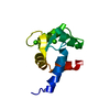
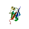
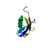
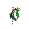
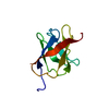
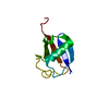
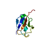
| Entry | Database: PDB / ID: 3b1l | ||||||
|---|---|---|---|---|---|---|---|
| Title | Crystal Structure of parkin ubiquitin-like domain R33Q mutant | ||||||
 Components Components | E3 ubiquitin-protein ligase parkin | ||||||
 Keywords Keywords |  LIGASE / Parkin / Ubiquitin / proteasome / Alfa-Beta-protein LIGASE / Parkin / Ubiquitin / proteasome / Alfa-Beta-protein | ||||||
| Function / homology |  Function and homology information Function and homology informationpositive regulation of neurotransmitter uptake / regulation protein catabolic process at presynapse / negative regulation of endoplasmic reticulum stress-induced neuron intrinsic apoptotic signaling pathway / mitochondrion-derived vesicle / negative regulation of spontaneous neurotransmitter secretion / negative regulation of intralumenal vesicle formation / PINK1-PRKN Mediated Mitophagy / negative regulation of exosomal secretion / Josephin domain DUBs / mitochondrion to lysosome vesicle-mediated transport ...positive regulation of neurotransmitter uptake / regulation protein catabolic process at presynapse / negative regulation of endoplasmic reticulum stress-induced neuron intrinsic apoptotic signaling pathway / mitochondrion-derived vesicle / negative regulation of spontaneous neurotransmitter secretion / negative regulation of intralumenal vesicle formation / PINK1-PRKN Mediated Mitophagy / negative regulation of exosomal secretion / Josephin domain DUBs / mitochondrion to lysosome vesicle-mediated transport / positive regulation of mitochondrial fusion / Regulation of necroptotic cell death / parkin-mediated stimulation of mitophagy in response to mitochondrial depolarization / cellular response to hydrogen sulfide / Aggrephagy / Parkin-FBXW7-Cul1 ubiquitin ligase complex / negative regulation of mitochondrial fusion / free ubiquitin chain polymerization / negative regulation of actin filament bundle assembly / RBR-type E3 ubiquitin transferase / positive regulation of protein linear polyubiquitination / F-box domain binding / negative regulation by host of viral genome replication / mitochondrion localization / positive regulation of mitophagy / mitochondrial fragmentation involved in apoptotic process / cellular response to toxic substance / regulation of dopamine metabolic process / dopaminergic synapse / regulation of cellular response to oxidative stress / negative regulation of excitatory postsynaptic potential / regulation of neurotransmitter secretion / cellular response to L-glutamate / autophagy of mitochondrion / negative regulation of intrinsic apoptotic signaling pathway by p53 class mediator / Antigen processing: Ubiquitination & Proteasome degradation / protein K6-linked ubiquitination / positive regulation of dendrite extension / norepinephrine metabolic process / protein localization to mitochondrion / negative regulation of oxidative stress-induced neuron intrinsic apoptotic signaling pathway / negative regulation of JNK cascade / positive regulation of protein localization to membrane / negative regulation of synaptic transmission, glutamatergic / protein K11-linked ubiquitination / protein metabolic process / synaptic transmission, dopaminergic / positive regulation of tumor necrosis factor-mediated signaling pathway / mitochondrial fission / aggresome assembly / ubiquitin conjugating enzyme binding / regulation of mitochondrion organization / aggresome / positive regulation of mitochondrial membrane potential / regulation of synaptic vesicle endocytosis / dopamine uptake involved in synaptic transmission / positive regulation of mitochondrial fission / dopamine metabolic process / intracellular vesicle / ubiquitin-specific protease binding / positive regulation of ATP biosynthetic process / startle response / negative regulation of release of cytochrome c from mitochondria / regulation of postsynaptic membrane neurotransmitter receptor levels / protein monoubiquitination / protein K63-linked ubiquitination / positive regulation of insulin secretion involved in cellular response to glucose stimulus / phospholipase binding / cullin family protein binding / mitophagy / negative regulation of mitochondrial fission / negative regulation of insulin secretion / protein autoubiquitination / protein K48-linked ubiquitination / negative regulation of endoplasmic reticulum stress-induced intrinsic apoptotic signaling pathway / negative regulation of reactive oxygen species biosynthetic process / ubiquitin ligase complex / Hsp70 protein binding / response to endoplasmic reticulum stress / mitochondrion organization / tubulin binding / adult locomotory behavior / negative regulation of protein phosphorylation / locomotory behavior / regulation of mitochondrial membrane potential / ubiquitin binding / learning / synaptic transmission, glutamatergic / regulation of autophagy / G protein-coupled receptor binding / PDZ domain binding / protein destabilization / protein catabolic process / modulation of chemical synaptic transmission / negative regulation of canonical Wnt signaling pathway / terminal bouton / synaptic vesicle membrane / beta-catenin binding / kinase binding / histone deacetylase bindingSimilarity search - Function | ||||||
| Biological species |  Mus musculus (house mouse) Mus musculus (house mouse) | ||||||
| Method | X-RAY DIFFRACTION / MOLECULAR REPLACEMENT / Resolution: 1.85 Å | ||||||
 Authors Authors | Tomoo, K. / Ikemiya, A. / Amami, Y. / In, Y. / Ishida, T. | ||||||
 Citation Citation | Journal: to be published Title: Crystal Structure of parkin ubiquitin-like domain R33Q mutant Authors: Tomoo, K. / Ikemiya, A. / Amami, Y. / In, Y. / Ishida, T. | ||||||
| History |
|
- Structure visualization
| Structure viewer | Molecule: MolmilJmol/JSmol |
|---|
- Downloads & links
-Download
| PDBx/mmCIF format | 3b1l.cif.gz | 27.2 KB | Display | PDBx/mmCIF format |
|---|---|---|---|---|
| PDB format | pdb3b1l.ent.gz | 17.9 KB | Display | PDB format |
| PDBx/mmJSON format | 3b1l.json.gz | Tree view | PDBx/mmJSON format | |
| Others |  Other downloads Other downloads |
-Validation report
| Arichive directory | https://data.pdbj.org/pub/pdb/validation_reports/b1/3b1lftp://data.pdbj.org/pub/pdb/validation_reports/b1/3b1l | HTTPS FTP |
|---|
-Related structure data
| Related structure data | |
|---|---|
| Similar structure data |


-Links
 PDBj
PDBj


- Assembly
| Deposited unit | 
| ||||||||
|---|---|---|---|---|---|---|---|---|---|
| 1 |
| ||||||||
| Unit cell |
|
-Components
| #1: Protein | Mass: 8700.982 Da / Num. of mol.: 1 / Fragment: Ubiquitin-like domain / Mutation: R33Q Source method: isolated from a genetically manipulated source Source: (gene. exp.) Mus musculus (house mouse) / Production host:  Escherichia coli (E. coli) Escherichia coli (E. coli)References: UniProt: Q9WVS6, Ligases; Forming carbon-nitrogen bonds; Acid-amino-acid ligases (peptide synthases) |
|---|---|
| #2: Water | ChemComp-HOH / Water Mass: 18.015 Da / Num. of mol.: 33 / Source method: isolated from a natural source / Formula: H2O Mass: 18.015 Da / Num. of mol.: 33 / Source method: isolated from a natural source / Formula: H2O |
-Experimental details
-Experiment
| Experiment | Method: X-RAY DIFFRACTION / Number of used crystals: 1 |
|---|
- Sample preparation
| Crystal | Density Matthews: 2.29 Å3/Da / Density % sol: 46.4 % |
|---|---|
| Crystal grow | Temperature: 291 K / Method: vapor diffusion, hanging drop / pH: 4.5 Details: 3M NaCl, 100mM acetate buffer, pH 4.5, VAPOR DIFFUSION, HANGING DROP, temperature 291.0K |
-Data collection
| Diffraction | Mean temperature: 100 K |
|---|---|
| Diffraction source | Source: ROTATING ANODE / Type: RIGAKU FR-E+ SUPERBRIGHT / Wavelength: 1.5418 Å |
| Radiation | Protocol: SINGLE WAVELENGTH / Monochromatic (M) / Laue (L): M / Scattering type: x-ray |
| Radiation wavelength | Wavelength: 1.5418 Å / Relative weight: 1 |
| Reflection | Resolution: 1.85→33.99 Å / Num. all: 6738 / Num. obs: 6718 / % possible obs: 99.7 % / Observed criterion σ(F): 2 / Observed criterion σ(I): 1 / Redundancy: 5.42 % / Rmerge(I) obs: 0.079 |
- Processing
| Software |
| |||||||||||||||||||||||||||||||||||||||||||||||||||||||||||||||||
|---|---|---|---|---|---|---|---|---|---|---|---|---|---|---|---|---|---|---|---|---|---|---|---|---|---|---|---|---|---|---|---|---|---|---|---|---|---|---|---|---|---|---|---|---|---|---|---|---|---|---|---|---|---|---|---|---|---|---|---|---|---|---|---|---|---|---|
| Refinement | Method to determine structure: MOLECULAR REPLACEMENT / Resolution: 1.85→25.36 Å / Cor.coef. Fo:Fc: 0.952 / Cor.coef. Fo:Fc free: 0.913 / Occupancy max: 1 / Occupancy min: 1 / SU B: 2.979 / SU ML: 0.09 / Cross valid method: THROUGHOUT / σ(F): 0 / ESU R Free: 0.145 / Stereochemistry target values: MAXIMUM LIKELIHOOD Details: HYDROGENS HAVE BEEN ADDED IN THE RIDING POSITIONS U VALUES: REFINED INDIVIDUALLY
| |||||||||||||||||||||||||||||||||||||||||||||||||||||||||||||||||
| Solvent computation | Ion probe radii: 0.8 Å / Shrinkage radii: 0.8 Å / VDW probe radii: 1.4 Å / Solvent model: MASK | |||||||||||||||||||||||||||||||||||||||||||||||||||||||||||||||||
| Displacement parameters | Biso max: 57.43 Å2 / Biso mean: 17.9307 Å2 / Biso min: 7.71 Å2
| |||||||||||||||||||||||||||||||||||||||||||||||||||||||||||||||||
| Refinement step | Cycle: LAST / Resolution: 1.85→25.36 Å
| |||||||||||||||||||||||||||||||||||||||||||||||||||||||||||||||||
| Refine LS restraints |
| |||||||||||||||||||||||||||||||||||||||||||||||||||||||||||||||||
| LS refinement shell | Resolution: 1.85→1.898 Å / Total num. of bins used: 20
|
