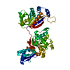
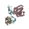
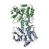
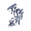
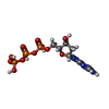
- PDB-3b0x: K263A mutant of PolX from Thermus thermophilus HB8 complexed with... -
+
Open data
ID or keywords:
Loading...
-
Basic information
Entry
Database: PDB / ID: 3b0x
Title
K263A mutant of PolX from Thermus thermophilus HB8 complexed with Ca-dGTP
Components
DNA polymerase beta family (X family)
Keywords
TRANSFERASE / Structural Genomics / RIKEN Structural Genomics/Proteomics Initiative / RSGI / POLXc / PHP / DNA polymerase / dRP lyase / 3'-5' exonuclease / AP endonuclease / DNA repair / nucleotide / DNA
Function / homology
Function and homology information
DNA-(apurinic or apyrimidinic site) lyase / DNA-directed DNA polymerase / DNA-directed DNA polymerase activity / DNA repair / DNA binding / metal ion binding / cytoplasm Similarity search - Function
: / DNA polymerase/3'-5' exonuclease PolX-like / PHP domain / PHP domain / Polymerase/histidinol phosphatase, N-terminal / DNA polymerase alpha chain like domain / Polymerase/histidinol phosphatase-like / RuvA domain 2-like / Helix-hairpin-helix domain / Beta Polymerase; domain 3 ...: / DNA polymerase/3'-5' exonuclease PolX-like / PHP domain / PHP domain / Polymerase/histidinol phosphatase, N-terminal / DNA polymerase alpha chain like domain / Polymerase/histidinol phosphatase-like / RuvA domain 2-like / Helix-hairpin-helix domain / Beta Polymerase; domain 3 / DNA polymerase, thumb domain / DNA polymerase family X, beta-like / DNA polymerase beta, N-terminal domain-like / Metal-dependent hydrolases / DNA-directed DNA polymerase X / DNA polymerase beta-like, N-terminal domain / Helix-hairpin-helix domain / DNA polymerase X family / DNA polymerase lambda lyase domain superfamily / DNA polymerase beta, thumb domain / DNA polymerase beta thumb / DNA polymerase, thumb domain superfamily / Beta Polymerase, domain 2 / Helix-hairpin-helix DNA-binding motif, class 1 / Helix-hairpin-helix DNA-binding motif class 1 / Beta Polymerase; domain 2 / Nucleotidyltransferase superfamily / 5' to 3' exonuclease, C-terminal subdomain / DNA polymerase; domain 1 / TIM Barrel / Alpha-Beta Barrel / 2-Layer Sandwich / Orthogonal Bundle / Mainly Alpha / Alpha Beta Similarity search - Domain/homology
In the structure databanks used in Yorodumi, some data are registered as the other names, "COVID-19 virus" and "2019-nCoV". Here are the details of the virus and the list of structure data.
Jan 31, 2019. EMDB accession codes are about to change! (news from PDBe EMDB page)
EMDB accession codes are about to change! (news from PDBe EMDB page)
The allocation of 4 digits for EMDB accession codes will soon come to an end. Whilst these codes will remain in use, new EMDB accession codes will include an additional digit and will expand incrementally as the available range of codes is exhausted. The current 4-digit format prefixed with “EMD-” (i.e. EMD-XXXX) will advance to a 5-digit format (i.e. EMD-XXXXX), and so on. It is currently estimated that the 4-digit codes will be depleted around Spring 2019, at which point the 5-digit format will come into force.
The EM Navigator/Yorodumi systems omit the EMD- prefix.
Related info.:Q: What is EMD? / ID/Accession-code notation in Yorodumi/EM Navigator
Yorodumi is a browser for structure data from EMDB, PDB, SASBDB, etc.
This page is also the successor to EM Navigator detail page, and also detail information page/front-end page for Omokage search.
The word "yorodu" (or yorozu) is an old Japanese word meaning "ten thousand". "mi" (miru) is to see.
Related info.:EMDB / PDB / SASBDB / Comparison of 3 databanks / Yorodumi Search / Aug 31, 2016. New EM Navigator & Yorodumi / Yorodumi Papers / Jmol/JSmol / Function and homology information / Changes in new EM Navigator and Yorodumi
 Movie
Movie Controller

 Yorodumi
 Open data
 Basic information
 Components
 Keywords
 TRANSFERASE /
  Function and homology information

 Authors
 Citation
 Structure visualization
 Downloads & links
 Other downloads
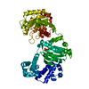


 PDBj
PDBj


 Assembly
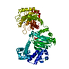


 Mass: 65.409 Da / Num. of mol.: 2 / Source method: obtained synthetically / Formula: Zn
 Mass: 40.078 Da / Num. of mol.: 4 / Source method: obtained synthetically / Formula: Ca
 Mass: 35.453 Da / Num. of mol.: 1 / Source method: obtained synthetically / Formula: Cl
 Mass: 507.181 Da / Num. of mol.: 1 / Source method: obtained synthetically / Formula: C10H16N5O13P3
 Sample preparation
 / Beamline: BL26B2 / Wavelength: 1 Å
 Processing