 Movie
Movie Controller


+ Open data

- Basic information
| Entry | Database: PDB / ID: 3asm | ||||||
|---|---|---|---|---|---|---|---|
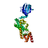
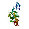
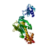
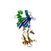
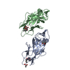
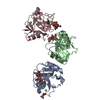
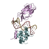
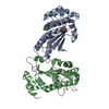
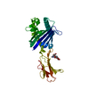
| Title | Crystal structure of Q54A mutant protein of Bst-RNase HIII | ||||||
 Components Components | Ribonuclease HIII | ||||||
 Keywords Keywords |  HYDROLASE / RNase / DNA/RNA binding HYDROLASE / RNase / DNA/RNA binding | ||||||
| Function / homology |  Function and homology information Function and homology informationRNA catabolic process / ribonuclease H / RNA-DNA hybrid ribonuclease activity / magnesium ion binding / RNA binding / cytoplasmSimilarity search - Function | ||||||
| Biological species |   Geobacillus stearothermophilus (bacteria) Geobacillus stearothermophilus (bacteria) | ||||||
| Method | X-RAY DIFFRACTION / SYNCHROTRON / MOLECULAR REPLACEMENT / Resolution: 2.603 Å | ||||||
 Authors Authors | Angkawidjaja, C. / Kanaya, S. | ||||||
 Citation Citation | Journal: Febs Lett. / Year: 2011 Title: Identification of the substrate binding site in the N-terminal TBP-like domain of RNase H3. Authors: Miyashita, S. / Tadokoro, T. / Angkawidjaja, C. / You, D.-J. / Koga, Y. / Takano, K. / Kanaya, S. | ||||||
| History |
|
- Structure visualization
| Structure viewer | Molecule: MolmilJmol/JSmol |
|---|
- Downloads & links
-Download
| PDBx/mmCIF format | 3asm.cif.gz | 71.7 KB | Display | PDBx/mmCIF format |
|---|---|---|---|---|
| PDB format | pdb3asm.ent.gz | 52.9 KB | Display | PDB format |
| PDBx/mmJSON format | 3asm.json.gz | Tree view | PDBx/mmJSON format | |
| Others |  Other downloads Other downloads |
-Validation report
| Arichive directory | https://data.pdbj.org/pub/pdb/validation_reports/as/3asmftp://data.pdbj.org/pub/pdb/validation_reports/as/3asm | HTTPS FTP |
|---|
-Related structure data
| Related structure data |  2d0aS S: Starting model for refinement |
|---|---|
| Similar structure data |

-Links
 PDBj
PDBj


- Assembly
| Deposited unit | 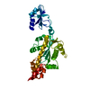
| ||||||||
|---|---|---|---|---|---|---|---|---|---|
| 1 |
| ||||||||
| Unit cell |
|
-Components
| #1: Protein | Mass: 33815.895 Da / Num. of mol.: 1 / Mutation: Q54A Source method: isolated from a genetically manipulated source Source: (gene. exp.) Geobacillus stearothermophilus (bacteria)Plasmid: pET25b(+) / Production host: Escherichia coli (E. coli) / Strain (production host): Mic2067(de3) / References: UniProt: Q6L6Q4, ribonuclease H |
|---|---|
| #2: Water | ChemComp-HOH / Water Mass: 18.015 Da / Num. of mol.: 58 / Source method: isolated from a natural source / Formula: H2O Mass: 18.015 Da / Num. of mol.: 58 / Source method: isolated from a natural source / Formula: H2O |
-Experimental details
-Experiment
| Experiment | Method: X-RAY DIFFRACTION / Number of used crystals: 1 |
|---|
- Sample preparation
| Crystal | Density Matthews: 2.62 Å3/Da / Density % sol: 53.08 % |
|---|---|
| Crystal grow | Temperature: 293 K / Method: vapor diffusion, sitting drop / pH: 8.5 Details: 0.085M Tris-HCl pH 8.5, 0.17M lithium sulfate, 25.5% PEG 4000, 15% glycerol, VAPOR DIFFUSION, SITTING DROP, temperature 293K |
-Data collection
| Diffraction | Mean temperature: 100 K |
|---|---|
| Diffraction source | Source: SYNCHROTRON / Site: SPring-8  / Beamline: BL38B1 / Wavelength: 1 Å / Beamline: BL38B1 / Wavelength: 1 Å |
| Detector | Type: RIGAKU JUPITER 210 / Detector: CCD / Date: Feb 14, 2008 / Details: mirrors |
| Radiation | Monochromator: mirros / Protocol: SINGLE WAVELENGTH / Monochromatic (M) / Laue (L): M / Scattering type: x-ray |
| Radiation wavelength | Wavelength: 1 Å / Relative weight: 1 |
| Reflection | Resolution: 2.6→50 Å / Num. obs: 11445 / % possible obs: 99.9 % / Observed criterion σ(F): 5 / Observed criterion σ(I): 5 / Redundancy: 6.9 % / Rmerge(I) obs: 0.106 / Rsym value: 0.086 / Net I/σ(I): 18.5 / Num. measured all: 240534 |
| Reflection shell | Resolution: 2.6→2.69 Å / Redundancy: 5.4 % / Rmerge(I) obs: 0.4 / Mean I/σ(I) obs: 3.33 / Num. unique all: 1102 / Rsym value: 0.355 / % possible all: 98.9 |
- Processing
| Software |
| |||||||||||||||||||||||||||||||||||||||||||||||||||||||||||||||||
|---|---|---|---|---|---|---|---|---|---|---|---|---|---|---|---|---|---|---|---|---|---|---|---|---|---|---|---|---|---|---|---|---|---|---|---|---|---|---|---|---|---|---|---|---|---|---|---|---|---|---|---|---|---|---|---|---|---|---|---|---|---|---|---|---|---|---|
| Refinement | Method to determine structure: MOLECULAR REPLACEMENT Starting model: PDB ENTRY 2D0A Resolution: 2.603→44.19 Å / Cor.coef. Fo:Fc: 0.925 / Cor.coef. Fo:Fc free: 0.905 / SU B: 8.81 / SU ML: 0.191 / Cross valid method: THROUGHOUT / ESU R Free: 0.29 Stereochemistry target values: MAXIMUM LIKELIHOOD WITH PHASES Details: HYDROGENS HAVE BEEN ADDED IN THE RIDING POSITIONS
| |||||||||||||||||||||||||||||||||||||||||||||||||||||||||||||||||
| Solvent computation | Ion probe radii: 0.8 Å / Shrinkage radii: 0.8 Å / VDW probe radii: 1.4 Å / Solvent model: MASK | |||||||||||||||||||||||||||||||||||||||||||||||||||||||||||||||||
| Displacement parameters | Biso mean: 25.873 Å2
| |||||||||||||||||||||||||||||||||||||||||||||||||||||||||||||||||
| Refinement step | Cycle: LAST / Resolution: 2.603→44.19 Å
| |||||||||||||||||||||||||||||||||||||||||||||||||||||||||||||||||
| Refine LS restraints |
| |||||||||||||||||||||||||||||||||||||||||||||||||||||||||||||||||
| LS refinement shell | Resolution: 2.603→2.67 Å / Total num. of bins used: 20
|
