 Movie
Movie Controller

[English] 日本語
 Yorodumi
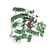
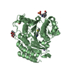
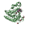
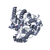
- PDB-3ani: Crystal structure of unsaturated glucuronyl hydrolase mutant D175... -
+ Open data

- Basic information
| Entry | Database: PDB / ID: 3ani | ||||||
|---|---|---|---|---|---|---|---|
| Title | Crystal structure of unsaturated glucuronyl hydrolase mutant D175N from Streptcoccus agalactiae | ||||||
 Components Components | Putative uncharacterized protein gbs1889 | ||||||
 Keywords Keywords |  HYDROLASE / alpha6/alpha6-barrel HYDROLASE / alpha6/alpha6-barrel | ||||||
| Function / homology |  Function and homology informationunsaturated chondroitin disaccharide hydrolase / unsaturated chondroitin disaccharide hydrolase activity / chondroitin hydrolase activity / polysaccharide catabolic process Function and homology informationunsaturated chondroitin disaccharide hydrolase / unsaturated chondroitin disaccharide hydrolase activity / chondroitin hydrolase activity / polysaccharide catabolic processSimilarity search - Function | ||||||
| Biological species |  Streptococcus agalactiae (bacteria) Streptococcus agalactiae (bacteria) | ||||||
| Method | X-RAY DIFFRACTION / SYNCHROTRON / MOLECULAR REPLACEMENT / Resolution: 2.5 Å | ||||||
 Authors Authors | Nakamichi, Y. / Maruyama, Y. / Mikami, B. / Hashimoto, W. / Murata, K. | ||||||
 Citation Citation | Journal: J.Biol.Chem. / Year: 2011 Title: Structural determinants in streptococcal unsaturated glucuronyl hydrolase for recognition of glycosaminoglycan sulfate groups Authors: Nakamichi, Y. / Maruyama, Y. / Mikami, B. / Hashimoto, W. / Murata, K. | ||||||
| History |
|
- Structure visualization
| Structure viewer | Molecule: MolmilJmol/JSmol |
|---|
- Downloads & links
-Download
| PDBx/mmCIF format | 3ani.cif.gz | 90.8 KB | Display | PDBx/mmCIF format |
|---|---|---|---|---|
| PDB format | pdb3ani.ent.gz | 69.2 KB | Display | PDB format |
| PDBx/mmJSON format | 3ani.json.gz | Tree view | PDBx/mmJSON format | |
| Others |  Other downloads Other downloads |
-Validation report
| Arichive directory | https://data.pdbj.org/pub/pdb/validation_reports/an/3aniftp://data.pdbj.org/pub/pdb/validation_reports/an/3ani | HTTPS FTP |
|---|
-Related structure data
| Related structure data |  3anjC  3ankC  2zzrS S: Starting model for refinement C: citing same article ( |
|---|---|
| Similar structure data |

-Links
 PDBj
PDBj
- Assembly
| Deposited unit | 
| ||||||||
|---|---|---|---|---|---|---|---|---|---|
| 1 |
| ||||||||
| 2 | 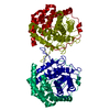
| ||||||||
| Unit cell |
|
-Components
| #1: Protein | Mass: 46640.691 Da / Num. of mol.: 1 / Mutation: D175N Source method: isolated from a genetically manipulated source Source: (gene. exp.) Streptococcus agalactiae (bacteria) / Strain: serogroup III/NEM316 / Gene: gbs1889, Ugl / Plasmid: pET21b / Production host: Escherichia coli (E. coli) / Strain (production host): HMS174References: UniProt: Q8E372, Hydrolases; Glycosylases; Glycosidases, i.e. enzymes that hydrolyse O- and S-glycosyl compounds |
|---|---|
| #2: Water | ChemComp-HOH / Water Mass: 18.015 Da / Num. of mol.: 61 / Source method: isolated from a natural source / Formula: H2O Mass: 18.015 Da / Num. of mol.: 61 / Source method: isolated from a natural source / Formula: H2O |
-Experimental details
-Experiment
| Experiment | Method: X-RAY DIFFRACTION / Number of used crystals: 1 |
|---|
- Sample preparation
| Crystal | Density Matthews: 2.1 Å3/Da / Density % sol: 41.33 % |
|---|---|
| Crystal grow | Temperature: 298 K / Method: vapor diffusion, sitting drop / pH: 7.5 Details: 40% ethylene glycol, 5% PEG 3000, 0.1M HEPES, pH 7.5, VAPOR DIFFUSION, SITTING DROP, temperature 298K |
-Data collection
| Diffraction | Mean temperature: 100 K |
|---|---|
| Diffraction source | Source: SYNCHROTRON / Site: SPring-8  / Beamline: BL38B1 / Wavelength: 1 Å / Beamline: BL38B1 / Wavelength: 1 Å |
| Detector | Type: ADSC QUANTUM 210 / Detector: CCD / Date: Jun 30, 2009 |
| Radiation | Monochromator: Fixed exit Si(111) double crystal monochromator Protocol: SINGLE WAVELENGTH / Monochromatic (M) / Laue (L): M / Scattering type: x-ray |
| Radiation wavelength | Wavelength: 1 Å / Relative weight: 1 |
| Reflection | Resolution: 2.5→30 Å / Num. obs: 13575 / % possible obs: 99 % / Redundancy: 3.7 % / Rmerge(I) obs: 0.064 |
| Reflection shell | Resolution: 2.5→2.59 Å / Redundancy: 3.8 % / Rmerge(I) obs: 0.352 / % possible all: 98.4 |
- Processing
| Software |
| ||||||||||||||||||||||||||||||||||||||||||||||||||||||||||||||||||||||||||||||||||||||||||||||||||||||||||||||||||||||||||||||||||||||||||||||||||||||||||||||||||||||||||
|---|---|---|---|---|---|---|---|---|---|---|---|---|---|---|---|---|---|---|---|---|---|---|---|---|---|---|---|---|---|---|---|---|---|---|---|---|---|---|---|---|---|---|---|---|---|---|---|---|---|---|---|---|---|---|---|---|---|---|---|---|---|---|---|---|---|---|---|---|---|---|---|---|---|---|---|---|---|---|---|---|---|---|---|---|---|---|---|---|---|---|---|---|---|---|---|---|---|---|---|---|---|---|---|---|---|---|---|---|---|---|---|---|---|---|---|---|---|---|---|---|---|---|---|---|---|---|---|---|---|---|---|---|---|---|---|---|---|---|---|---|---|---|---|---|---|---|---|---|---|---|---|---|---|---|---|---|---|---|---|---|---|---|---|---|---|---|---|---|---|---|---|
| Refinement | Method to determine structure: MOLECULAR REPLACEMENT Starting model: 2ZZR Resolution: 2.5→29.16 Å / Cor.coef. Fo:Fc: 0.942 / Cor.coef. Fo:Fc free: 0.883 / SU B: 9.38 / SU ML: 0.215 / Cross valid method: THROUGHOUT / ESU R Free: 0.336 / Stereochemistry target values: MAXIMUM LIKELIHOOD / Details: HYDROGENS HAVE BEEN ADDED IN THE RIDING POSITIONS
| ||||||||||||||||||||||||||||||||||||||||||||||||||||||||||||||||||||||||||||||||||||||||||||||||||||||||||||||||||||||||||||||||||||||||||||||||||||||||||||||||||||||||||
| Solvent computation | Ion probe radii: 0.8 Å / Shrinkage radii: 0.8 Å / VDW probe radii: 1.4 Å / Solvent model: MASK | ||||||||||||||||||||||||||||||||||||||||||||||||||||||||||||||||||||||||||||||||||||||||||||||||||||||||||||||||||||||||||||||||||||||||||||||||||||||||||||||||||||||||||
| Displacement parameters | Biso mean: 35.806 Å2
| ||||||||||||||||||||||||||||||||||||||||||||||||||||||||||||||||||||||||||||||||||||||||||||||||||||||||||||||||||||||||||||||||||||||||||||||||||||||||||||||||||||||||||
| Refinement step | Cycle: LAST / Resolution: 2.5→29.16 Å
| ||||||||||||||||||||||||||||||||||||||||||||||||||||||||||||||||||||||||||||||||||||||||||||||||||||||||||||||||||||||||||||||||||||||||||||||||||||||||||||||||||||||||||
| Refine LS restraints |
| ||||||||||||||||||||||||||||||||||||||||||||||||||||||||||||||||||||||||||||||||||||||||||||||||||||||||||||||||||||||||||||||||||||||||||||||||||||||||||||||||||||||||||
| LS refinement shell | Resolution: 2.499→2.564 Å / Total num. of bins used: 20
|
