 Movie
Movie Controller

[English] 日本語
 Yorodumi
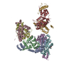
- PDB-3alz: Crystal structure of the measles virus hemagglutinin bound to its... -
+ Open data

- Basic information
| Entry | Database: PDB / ID: 3alz | ||||||
|---|---|---|---|---|---|---|---|
| Title | Crystal structure of the measles virus hemagglutinin bound to its cellular receptor SLAM (Form I) | ||||||
 Components Components |
| ||||||
 Keywords Keywords |  viral protein/membrane protein / viral protein-receptor complex / six-bladed beta-propeller fold / Immunoglobulin fold / beta-sandwich / viral protein-membrane protein complex viral protein/membrane protein / viral protein-receptor complex / six-bladed beta-propeller fold / Immunoglobulin fold / beta-sandwich / viral protein-membrane protein complex | ||||||
| Function / homology |  Function and homology informationlymphocyte activation / host cell membrane / positive regulation of type II interferon production / signaling receptor activity / host cell surface receptor binding / symbiont entry into host cell / external side of plasma membrane / viral envelope / virion attachment to host cell / host cell plasma membrane ...lymphocyte activation / host cell membrane / positive regulation of type II interferon production / signaling receptor activity / host cell surface receptor binding / symbiont entry into host cell / external side of plasma membrane / viral envelope / virion attachment to host cell / host cell plasma membrane / virion membrane / membrane Function and homology informationlymphocyte activation / host cell membrane / positive regulation of type II interferon production / signaling receptor activity / host cell surface receptor binding / symbiont entry into host cell / external side of plasma membrane / viral envelope / virion attachment to host cell / host cell plasma membrane ...lymphocyte activation / host cell membrane / positive regulation of type II interferon production / signaling receptor activity / host cell surface receptor binding / symbiont entry into host cell / external side of plasma membrane / viral envelope / virion attachment to host cell / host cell plasma membrane / virion membrane / membraneSimilarity search - Function | ||||||
| Biological species |   Measles virus Measles virus Saguinus oedipus (cotton-top tamarin) Saguinus oedipus (cotton-top tamarin) | ||||||
| Method | X-RAY DIFFRACTION / SYNCHROTRON / MOLECULAR REPLACEMENT / Resolution: 4.515 Å | ||||||
 Authors Authors | Hashiguchi, T. / Ose, T. / Kubota, M. / Maita, N. / Kamishikiryo, J. / Maenaka, K. / Yanagi, Y. | ||||||
 Citation Citation | Journal: Nat.Struct.Mol.Biol. / Year: 2011 Title: Structure of the measles virus hemagglutinin bound to its cellular receptor SLAM Authors: Hashiguchi, T. / Ose, T. / Kubota, M. / Maita, N. / Kamishikiryo, J. / Maenaka, K. / Yanagi, Y. | ||||||
| History |
|
- Structure visualization
| Structure viewer | Molecule: MolmilJmol/JSmol |
|---|
- Downloads & links
-Download
| PDBx/mmCIF format | 3alz.cif.gz | 235.8 KB | Display | PDBx/mmCIF format |
|---|---|---|---|---|
| PDB format | pdb3alz.ent.gz | 190.5 KB | Display | PDB format |
| PDBx/mmJSON format | 3alz.json.gz | Tree view | PDBx/mmJSON format | |
| Others |  Other downloads Other downloads |
-Validation report
| Arichive directory | https://data.pdbj.org/pub/pdb/validation_reports/al/3alzftp://data.pdbj.org/pub/pdb/validation_reports/al/3alz | HTTPS FTP |
|---|
-Related structure data
| Related structure data |  3alwC 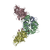 3alxC 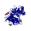 2zb6S C: citing same article ( S: Starting model for refinement |
|---|---|
| Similar structure data |


-Links
 PDBj
PDBj

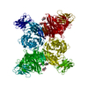
- Assembly
| Deposited unit | 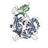
| ||||||||
|---|---|---|---|---|---|---|---|---|---|
| 1 | 
| ||||||||
| 2 | x 6
| ||||||||
| Unit cell |
|
-Components
| #1: Protein | Mass: 53423.656 Da / Num. of mol.: 1 / Fragment: head domain Source method: isolated from a genetically manipulated source Source: (gene. exp.) Measles virus / Strain: Edmonston B / Gene: Hemagglutinin / Plasmid: pCA7 / Cell line (production host): HEK293SGnTI(-) / Production host:  Homo sapiens (human) / References: UniProt: E2RZS2, UniProt: P08362*PLUS Homo sapiens (human) / References: UniProt: E2RZS2, UniProt: P08362*PLUS |
|---|---|
| #2: Protein | Mass: 17245.938 Da / Num. of mol.: 1 / Fragment: V domain, residues 1-140 Source method: isolated from a genetically manipulated source Source: (gene. exp.) Saguinus oedipus (cotton-top tamarin) / Strain: B95a / Gene: SLAM (CD150) / Plasmid: pCA7 / Cell line (production host): HEK293SGnTI(-) / Production host: Homo sapiens (human) / References: UniProt: Q9GJT3 |
| #3: Sugar | N-Acetylglucosamine  Type: D-saccharide, beta linking / Mass: 221.208 Da / Num. of mol.: 2 Type: D-saccharide, beta linking / Mass: 221.208 Da / Num. of mol.: 2Source method: isolated from a genetically manipulated source Formula: C8H15NO6 |
-Experimental details
-Experiment
| Experiment | Method: X-RAY DIFFRACTION / Number of used crystals: 1 |
|---|
- Sample preparation
| Crystal | Density Matthews: 8.055825 Å3/Da / Density % sol: 84.731544 % |
|---|---|
| Crystal grow | Temperature: 293 K / Method: vapor diffusion, sitting drop / pH: 6.5 Details: 0.1M MES pH6.5, 0.75M Li2SO4, VAPOR DIFFUSION, SITTING DROP, temperature 293K |
-Data collection
| Diffraction | Mean temperature: 100 K |
|---|---|
| Diffraction source | Source: SYNCHROTRON / Site: Photon Factory  / Beamline: BL-17A / Wavelength: 1 Å / Beamline: BL-17A / Wavelength: 1 Å |
| Detector | Type: ADSC QUANTUM 270 / Detector: CCD / Date: Mar 10, 2009 |
| Radiation | Protocol: SINGLE WAVELENGTH / Monochromatic (M) / Laue (L): M / Scattering type: x-ray |
| Radiation wavelength | Wavelength: 1 Å / Relative weight: 1 |
| Reflection | Resolution: 4.5→30.04 Å / Num. obs: 14397 / % possible obs: 100 % / Redundancy: 15.5 % / Biso Wilson estimate: 154.7 Å2 / Rmerge(I) obs: 0.078 / Net I/σ(I): 15.4 |
| Reflection shell | Resolution: 4.5→4.66 Å / Redundancy: 10.9 % / Rmerge(I) obs: 0.592 / Mean I/σ(I) obs: 4.9 / Num. unique all: 1396 / % possible all: 100 |
- Processing
| Software |
| |||||||||||||||||||||||||||||||||||||||||||||||||||||||||||||||||||||||||||
|---|---|---|---|---|---|---|---|---|---|---|---|---|---|---|---|---|---|---|---|---|---|---|---|---|---|---|---|---|---|---|---|---|---|---|---|---|---|---|---|---|---|---|---|---|---|---|---|---|---|---|---|---|---|---|---|---|---|---|---|---|---|---|---|---|---|---|---|---|---|---|---|---|---|---|---|---|
| Refinement | Method to determine structure: MOLECULAR REPLACEMENT Starting model: PDB ENTRY 2ZB6 Resolution: 4.515→30.039 Å / Occupancy max: 1 / Occupancy min: 1 / FOM work R set: 0.7401 / SU ML: 0.72 / σ(F): 1.34 / Phase error: 31.44 / Stereochemistry target values: ML Details: DUE TO LOW RESOLUTION, THE REFINEMENT PROTOCOL WAS LIMITED TO THE FOLLOWING THREE STEPS AFTER MOLECULAR REPLACEMENT. 1. RIGID BODY REFINEMENT OF INDIVIDUAL DOMAINS 2. TLS REFINEMENT OF ...Details: DUE TO LOW RESOLUTION, THE REFINEMENT PROTOCOL WAS LIMITED TO THE FOLLOWING THREE STEPS AFTER MOLECULAR REPLACEMENT. 1. RIGID BODY REFINEMENT OF INDIVIDUAL DOMAINS 2. TLS REFINEMENT OF SELECTED PORTIONS 3. STRUCTURE REGULARIZATION TO AVOID MINOR CLASHES. WE PERFORMED NO INDIVIDUAL ATOM REFINEMENT.
| |||||||||||||||||||||||||||||||||||||||||||||||||||||||||||||||||||||||||||
| Solvent computation | Shrinkage radii: 0.83 Å / VDW probe radii: 1.1 Å / Solvent model: FLAT BULK SOLVENT MODEL / Bsol: 199.342 Å2 / ksol: 0.301 e/Å3 | |||||||||||||||||||||||||||||||||||||||||||||||||||||||||||||||||||||||||||
| Displacement parameters | Biso max: 712.58 Å2 / Biso mean: 271.8932 Å2 / Biso min: 128.63 Å2
| |||||||||||||||||||||||||||||||||||||||||||||||||||||||||||||||||||||||||||
| Refine analyze |
| |||||||||||||||||||||||||||||||||||||||||||||||||||||||||||||||||||||||||||
| Refinement step | Cycle: LAST / Resolution: 4.515→30.039 Å
| |||||||||||||||||||||||||||||||||||||||||||||||||||||||||||||||||||||||||||
| Refine LS restraints |
| |||||||||||||||||||||||||||||||||||||||||||||||||||||||||||||||||||||||||||
| LS refinement shell |
| |||||||||||||||||||||||||||||||||||||||||||||||||||||||||||||||||||||||||||
| Refinement TLS params. | Method: refined / Refine-ID: X-RAY DIFFRACTION
| |||||||||||||||||||||||||||||||||||||||||||||||||||||||||||||||||||||||||||
| Refinement TLS group |
|
