 Movie
Movie Controller

[English] 日本語
 Yorodumi
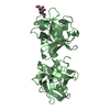
- PDB-3aj5: HA1 (HA33) subcomponent of botulinum type C progenitor toxin comp... -
+ Open data

- Basic information
| Entry | Database: PDB / ID: 3aj5 | ||||||
|---|---|---|---|---|---|---|---|
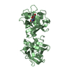
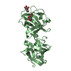
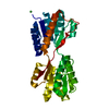
| Title | HA1 (HA33) subcomponent of botulinum type C progenitor toxin complexed with N-acetylgalactosamine, bound at site II | ||||||
 Components Components | Main hemagglutinin component | ||||||
 Keywords Keywords |  TOXIN / beta-trefoil / hemagglutinin TOXIN / beta-trefoil / hemagglutinin | ||||||
| Function / homology |  Function and homology information Function and homology information | ||||||
| Biological species |   Clostridium botulinum (bacteria) Clostridium botulinum (bacteria) | ||||||
| Method | X-RAY DIFFRACTION / SYNCHROTRON / MOLECULAR REPLACEMENT / Resolution: 1.8 Å | ||||||
 Authors Authors | Nakamura, T. / Tonozuka, T. / Sato, R. / Oguma, K. / Nishikawa, A. | ||||||
 Citation Citation | Journal: Arch.Biochem.Biophys. / Year: 2011 Title: Molecular diversity of the two sugar-binding sites of the beta-trefoil lectin HA33/C (HA1) from Clostridium botulinum type C neurotoxin Authors: Nakamura, T. / Tonozuka, T. / Ito, S. / Takeda, Y. / Sato, R. / Matsuo, I. / Ito, Y. / Oguma, K. / Nishikawa, A. #1: Journal: J.Mol.Biol. / Year: 2008Title: Sugar-binding sites of the HA1 subcomponent of Clostridium botulinum type C progenitor toxin Authors: Nakamura, T. / Tonozuka, T. / Ide, A. / Yuzawa, T. / Oguma, K. / Nishikawa, A. | ||||||
| History |
|
- Structure visualization
| Structure viewer | Molecule: MolmilJmol/JSmol |
|---|
- Downloads & links
-Download
| PDBx/mmCIF format | 3aj5.cif.gz | 145.9 KB | Display | PDBx/mmCIF format |
|---|---|---|---|---|
| PDB format | pdb3aj5.ent.gz | 114 KB | Display | PDB format |
| PDBx/mmJSON format | 3aj5.json.gz | Tree view | PDBx/mmJSON format | |
| Others |  Other downloads Other downloads |
-Validation report
| Arichive directory | https://data.pdbj.org/pub/pdb/validation_reports/aj/3aj5ftp://data.pdbj.org/pub/pdb/validation_reports/aj/3aj5 | HTTPS FTP |
|---|
-Related structure data
| Related structure data | 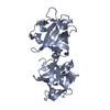 3aj6C 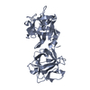 3ah2S S: Starting model for refinement C: citing same article ( |
|---|---|
| Similar structure data |


-Links
 PDBj
PDBj


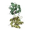
- Assembly
| Deposited unit | 
| ||||||||
|---|---|---|---|---|---|---|---|---|---|
| 1 | 
| ||||||||
| 2 | 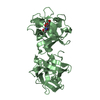
| ||||||||
| 3 |
| ||||||||
| Unit cell |
|
-Components
| #1: Protein | Mass: 33779.488 Da / Num. of mol.: 2 Source method: isolated from a genetically manipulated source Source: (gene. exp.) Clostridium botulinum (bacteria) / Strain: type C / Plasmid: pMAL-2X FLAG-HA1 / Production host: Escherichia coli (E. coli) / Strain (production host): JM109 / References: UniProt: P46084, UniProt: P0DPR0*PLUS#2: Sugar | N-Acetylgalactosamine  Type: D-saccharide, beta linking / Mass: 221.208 Da / Num. of mol.: 2 Type: D-saccharide, beta linking / Mass: 221.208 Da / Num. of mol.: 2Source method: isolated from a genetically manipulated source Formula: C8H15NO6 #3: Water | ChemComp-HOH / | Water Mass: 18.015 Da / Num. of mol.: 729 / Source method: isolated from a natural source / Formula: H2O Mass: 18.015 Da / Num. of mol.: 729 / Source method: isolated from a natural source / Formula: H2O |
|---|
-Experimental details
-Experiment
| Experiment | Method: X-RAY DIFFRACTION / Number of used crystals: 1 |
|---|
- Sample preparation
| Crystal | Density Matthews: 2.54 Å3/Da / Density % sol: 51.59 % |
|---|---|
| Crystal grow | Temperature: 293 K / Method: vapor diffusion, hanging drop / pH: 6 Details: 12% ethanol, 1.7M sodium chloride, pH 6.0, VAPOR DIFFUSION, HANGING DROP, temperature 293K |
-Data collection
| Diffraction | Mean temperature: 100 K |
|---|---|
| Diffraction source | Source: SYNCHROTRON / Site: Photon Factory  / Beamline: AR-NW12A / Wavelength: 1 Å / Beamline: AR-NW12A / Wavelength: 1 Å |
| Detector | Type: ADSC QUANTUM 210 / Detector: CCD / Date: Mar 9, 2008 |
| Radiation | Protocol: SINGLE WAVELENGTH / Monochromatic (M) / Laue (L): M / Scattering type: x-ray |
| Radiation wavelength | Wavelength: 1 Å / Relative weight: 1 |
| Reflection | Resolution: 1.8→50 Å / Num. obs: 62088 / % possible obs: 98.6 % / Redundancy: 3.8 % / Rmerge(I) obs: 0.071 / Net I/σ(I): 18.3 |
| Reflection shell | Resolution: 1.8→1.86 Å / Redundancy: 3.8 % / Rmerge(I) obs: 0.4 / Mean I/σ(I) obs: 2.8 / Num. unique all: 6068 / % possible all: 97.5 |
- Processing
| Software |
| ||||||||||||||||||||||||||||||||||||||||||||||||||||||||||||||||||||||||||||||||||||||||||||||||||||||||||||||||||||||||||||||||||||||||||||||||||||||||||||||||||||||||||
|---|---|---|---|---|---|---|---|---|---|---|---|---|---|---|---|---|---|---|---|---|---|---|---|---|---|---|---|---|---|---|---|---|---|---|---|---|---|---|---|---|---|---|---|---|---|---|---|---|---|---|---|---|---|---|---|---|---|---|---|---|---|---|---|---|---|---|---|---|---|---|---|---|---|---|---|---|---|---|---|---|---|---|---|---|---|---|---|---|---|---|---|---|---|---|---|---|---|---|---|---|---|---|---|---|---|---|---|---|---|---|---|---|---|---|---|---|---|---|---|---|---|---|---|---|---|---|---|---|---|---|---|---|---|---|---|---|---|---|---|---|---|---|---|---|---|---|---|---|---|---|---|---|---|---|---|---|---|---|---|---|---|---|---|---|---|---|---|---|---|---|---|
| Refinement | Method to determine structure: MOLECULAR REPLACEMENT Starting model: 3AH2 Resolution: 1.8→31 Å / Cor.coef. Fo:Fc: 0.961 / Cor.coef. Fo:Fc free: 0.94 / Cross valid method: THROUGHOUT / ESU R Free: 0.114 / Stereochemistry target values: MAXIMUM LIKELIHOOD / Details: HYDROGENS HAVE BEEN ADDED IN THE RIDING POSITIONS
| ||||||||||||||||||||||||||||||||||||||||||||||||||||||||||||||||||||||||||||||||||||||||||||||||||||||||||||||||||||||||||||||||||||||||||||||||||||||||||||||||||||||||||
| Solvent computation | Ion probe radii: 0.8 Å / Shrinkage radii: 0.8 Å / VDW probe radii: 1.4 Å / Solvent model: BABINET MODEL WITH MASK | ||||||||||||||||||||||||||||||||||||||||||||||||||||||||||||||||||||||||||||||||||||||||||||||||||||||||||||||||||||||||||||||||||||||||||||||||||||||||||||||||||||||||||
| Displacement parameters | Biso mean: 18.23 Å2
| ||||||||||||||||||||||||||||||||||||||||||||||||||||||||||||||||||||||||||||||||||||||||||||||||||||||||||||||||||||||||||||||||||||||||||||||||||||||||||||||||||||||||||
| Refinement step | Cycle: LAST / Resolution: 1.8→31 Å
| ||||||||||||||||||||||||||||||||||||||||||||||||||||||||||||||||||||||||||||||||||||||||||||||||||||||||||||||||||||||||||||||||||||||||||||||||||||||||||||||||||||||||||
| Refine LS restraints |
| ||||||||||||||||||||||||||||||||||||||||||||||||||||||||||||||||||||||||||||||||||||||||||||||||||||||||||||||||||||||||||||||||||||||||||||||||||||||||||||||||||||||||||
| LS refinement shell | Resolution: 1.799→1.846 Å / Total num. of bins used: 20
|
