 Movie
Movie Controller


+ Open data

- Basic information
| Entry | Database: PDB / ID: 3a7k | ||||||
|---|---|---|---|---|---|---|---|
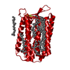
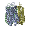
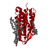
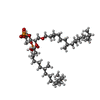
| Title | Crystal structure of halorhodopsin from Natronomonas pharaonis | ||||||
 Components Components | Halorhodopsin | ||||||
 Keywords Keywords | MEMBRANE PROTEIN / Light-driven chloride ion pump / Trimeric Bacterioruberin-Protein Complex / Retinal Protein | ||||||
| Function / homology |  Function and homology information Function and homology informationRhopdopsin 7-helix transmembrane proteins / Rhodopsin 7-helix transmembrane proteins / Up-down Bundle / Mainly Alpha Similarity search - Domain/homology | ||||||
| Biological species |  Natronomonas pharaonis DSM 2160 (archaea) Natronomonas pharaonis DSM 2160 (archaea) | ||||||
| Method | X-RAY DIFFRACTION / SYNCHROTRON / MOLECULAR REPLACEMENT / Resolution: 2 Å | ||||||
 Authors Authors | Kouyama, T. | ||||||
 Citation Citation | Journal: J.Mol.Biol. / Year: 2010 Title: Crystal Structure of the Light-Driven Chloride Pump Halorhodopsin from Natronomonas pharaonis. Authors: Kouyama, T. / Kanada, S. / Takeguchi, Y. / Narusawa, A. / Murakami, M. / Ihara, K. | ||||||
| History |
|
- Structure visualization
| Structure viewer | Molecule: MolmilJmol/JSmol |
|---|
- Downloads & links
-Download
| PDBx/mmCIF format | 3a7k.cif.gz | 183.9 KB | Display | PDBx/mmCIF format |
|---|---|---|---|---|
| PDB format | pdb3a7k.ent.gz | 141.5 KB | Display | PDB format |
| PDBx/mmJSON format | 3a7k.json.gz | Tree view | PDBx/mmJSON format | |
| Others |  Other downloads Other downloads |
-Validation report
| Arichive directory | https://data.pdbj.org/pub/pdb/validation_reports/a7/3a7kftp://data.pdbj.org/pub/pdb/validation_reports/a7/3a7k | HTTPS FTP |
|---|
-Related structure data
| Related structure data | 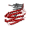 1e12S  3a4b S: Starting model for refinement |
|---|---|
| Similar structure data |


-Links
 PDBj
PDBj


- Assembly
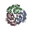
| Deposited unit | 
| ||||||||
|---|---|---|---|---|---|---|---|---|---|
| 1 |
| ||||||||
| Unit cell |
|
-Components
-Protein , 1 types, 3 molecules ABD
| #1: Protein | Mass: 30975.096 Da / Num. of mol.: 3 / Source method: isolated from a natural source Details: STRAIN MK-1 WAS A HALORHODOPSIN-OVERPRODUCING MUTANT GENERATED FROM TYPE STRAIN D2160T. Source: (natural) Natronomonas pharaonis DSM 2160 (archaea) / References: UniProt: Q3ITX1 |
|---|
-Non-polymers , 7 types, 174 molecules 
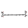
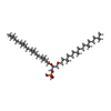
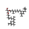


| #2: Chemical | Retinal Mass: 284.436 Da / Num. of mol.: 3 / Source method: obtained synthetically / Formula: C20H28O Mass: 284.436 Da / Num. of mol.: 3 / Source method: obtained synthetically / Formula: C20H28O#3: Chemical | Halobacterium Mass: 741.136 Da / Num. of mol.: 3 / Source method: obtained synthetically / Formula: C50H76O4 Mass: 741.136 Da / Num. of mol.: 3 / Source method: obtained synthetically / Formula: C50H76O4#4: Chemical |  Mass: 733.137 Da / Num. of mol.: 3 / Source method: obtained synthetically / Formula: C43H89O6P Mass: 733.137 Da / Num. of mol.: 3 / Source method: obtained synthetically / Formula: C43H89O6P#5: Chemical | ChemComp-L2P /  Mass: 653.157 Da / Num. of mol.: 6 / Source method: obtained synthetically / Formula: C43H88O3 Mass: 653.157 Da / Num. of mol.: 6 / Source method: obtained synthetically / Formula: C43H88O3#6: Chemical | ChemComp-L3P /  Mass: 885.179 Da / Num. of mol.: 9 / Source method: obtained synthetically / Formula: C46H94O11P2 Mass: 885.179 Da / Num. of mol.: 9 / Source method: obtained synthetically / Formula: C46H94O11P2#7: Chemical | ChemComp-CL / Chloride Mass: 35.453 Da / Num. of mol.: 6 / Source method: obtained synthetically / Formula: Cl Mass: 35.453 Da / Num. of mol.: 6 / Source method: obtained synthetically / Formula: Cl#8: Water | ChemComp-HOH / | WaterMass: 18.015 Da / Num. of mol.: 144 / Source method: isolated from a natural source / Formula: H2O |
|---|
-Experimental details
-Experiment
| Experiment | Method: X-RAY DIFFRACTION / Number of used crystals: 1 |
|---|
- Sample preparation
| Crystal | Density Matthews: 3.2 Å3/Da / Density % sol: 61.59 % |
|---|---|
| Crystal grow | Temperature: 293 K / Method: vapor diffusion, sitting drop / pH: 8 Details: A DROP CONTAINING 1MG/ML PROTEIN, 1M AMMONIUM SULFATE, 0.16M NACL, 0.04M TRIS-HCL, 0.04% NAN2, 5MG/ML NONYL GLUCOSIDE WAS CONCENTRATED BY VAPOR DIFFUSION AGAINST A RESERVE SOLUTION ...Details: A DROP CONTAINING 1MG/ML PROTEIN, 1M AMMONIUM SULFATE, 0.16M NACL, 0.04M TRIS-HCL, 0.04% NAN2, 5MG/ML NONYL GLUCOSIDE WAS CONCENTRATED BY VAPOR DIFFUSION AGAINST A RESERVE SOLUTION CONTAINING 2.6 M AMMONIUM SULFATE, 0.16M NACL, 0.04M TRIS-HCL, PH 8, VAPOR DIFFUSION, SITTING DROP, TEMPERATURE 293K |
-Data collection
| Diffraction | Mean temperature: 100 K |
|---|---|
| Diffraction source | Source: SYNCHROTRON / Site: SPring-8  / Beamline: BL41XU / Wavelength: 1 Å / Beamline: BL41XU / Wavelength: 1 Å |
| Detector | Type: MAR CCD 165 mm / Detector: CCD / Date: Jan 1, 2009 / Details: mirrors |
| Radiation | Monochromator: GRAPHITE / Protocol: SINGLE WAVELENGTH / Monochromatic (M) / Laue (L): M / Scattering type: x-ray |
| Radiation wavelength | Wavelength: 1 Å / Relative weight: 1 |
| Reflection | Resolution: 2→60.1 Å / Num. obs: 79092 / % possible obs: 99.5 % / Observed criterion σ(F): 0 / Observed criterion σ(I): 0 / Redundancy: 2.7 % / Biso Wilson estimate: 31.2 Å2 / Rmerge(I) obs: 0.072 / Rsym value: 0.072 / Net I/σ(I): 9.9 |
| Reflection shell | Resolution: 2→2.11 Å / Redundancy: 2.7 % / Rmerge(I) obs: 0.556 / Mean I/σ(I) obs: 2 / Rsym value: 0.556 / % possible all: 100 |
- Processing
| Software |
| ||||||||||||||||||||||||||||||||||||||||||
|---|---|---|---|---|---|---|---|---|---|---|---|---|---|---|---|---|---|---|---|---|---|---|---|---|---|---|---|---|---|---|---|---|---|---|---|---|---|---|---|---|---|---|---|
| Refinement | Method to determine structure: MOLECULAR REPLACEMENT Starting model: PDB ENTRY 1e12 Resolution: 2→15 Å / Isotropic thermal model: Isotropic / Cross valid method: THROUGHOUT / σ(F): 0 / σ(I): 0 / Stereochemistry target values: Engh & Huber
| ||||||||||||||||||||||||||||||||||||||||||
| Solvent computation | Bsol: 109.866 Å2 | ||||||||||||||||||||||||||||||||||||||||||
| Displacement parameters | Biso mean: 49.222 Å2
| ||||||||||||||||||||||||||||||||||||||||||
| Refine analyze |
| ||||||||||||||||||||||||||||||||||||||||||
| Refinement step | Cycle: LAST / Resolution: 2→15 Å
| ||||||||||||||||||||||||||||||||||||||||||
| Refine LS restraints |
| ||||||||||||||||||||||||||||||||||||||||||
| LS refinement shell |
| ||||||||||||||||||||||||||||||||||||||||||
| Xplor file |
|
