 Movie
Movie Controller

[English] 日本語
 Yorodumi
- PDB-2zr8: Crystal Structure of Modified Serine Racemase complexed with Serine -
+ Open data

- Basic information
| Entry | Database: PDB / ID: 2zr8 | ||||||
|---|---|---|---|---|---|---|---|
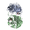
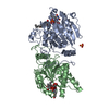
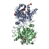
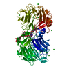
| Title | Crystal Structure of Modified Serine Racemase complexed with Serine | ||||||
 Components Components | Uncharacterized protein C320.14 | ||||||
 Keywords Keywords |  ISOMERASE / PLP-dependent / Lyase / Pyridoxal phosphate ISOMERASE / PLP-dependent / Lyase / Pyridoxal phosphate | ||||||
| Function / homology |  Function and homology information Function and homology informationSerine biosynthesis / serine racemase / threonine racemase activity / serine racemase activity / D-serine ammonia-lyase / D-serine ammonia-lyase activity / L-serine ammonia-lyase / L-serine ammonia-lyase activity / D-serine metabolic process / L-serine metabolic process ...Serine biosynthesis / serine racemase / threonine racemase activity / serine racemase activity / D-serine ammonia-lyase / D-serine ammonia-lyase activity / L-serine ammonia-lyase / L-serine ammonia-lyase activity / D-serine metabolic process / L-serine metabolic process / pyridoxal phosphate binding / magnesium ion binding / protein homodimerization activity / ATP bindingSimilarity search - Function | ||||||
| Biological species |  Schizosaccharomyces pombe (fission yeast) Schizosaccharomyces pombe (fission yeast) | ||||||
| Method | X-RAY DIFFRACTION / SYNCHROTRON / MOLECULAR REPLACEMENT / Resolution: 2.2 Å | ||||||
 Authors Authors | Goto, M. | ||||||
 Citation Citation | Journal: J.Biol.Chem. / Year: 2009 Title: Crystal structure of a homolog of mammalian serine racemase from Schizosaccharomyces pombe Authors: Goto, M. / Yamauchi, T. / Kamiya, N. / Miyahara, I. / Yoshimura, T. / Mihara, H. / Kurihara, T. / Hirotsu, K. / Esaki, N. | ||||||
| History |
|
- Structure visualization
| Structure viewer | Molecule: MolmilJmol/JSmol |
|---|
- Downloads & links
-Download
| PDBx/mmCIF format | 2zr8.cif.gz | 77 KB | Display | PDBx/mmCIF format |
|---|---|---|---|---|
| PDB format | pdb2zr8.ent.gz | 56.1 KB | Display | PDB format |
| PDBx/mmJSON format | 2zr8.json.gz | Tree view | PDBx/mmJSON format | |
| Others |  Other downloads Other downloads |
-Validation report
| Arichive directory | https://data.pdbj.org/pub/pdb/validation_reports/zr/2zr8ftp://data.pdbj.org/pub/pdb/validation_reports/zr/2zr8 | HTTPS FTP |
|---|
-Related structure data
| Related structure data | 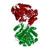 1wtcC 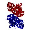 2zpuS S: Starting model for refinement C: citing same article ( |
|---|---|
| Similar structure data |


-Links
 PDBj
PDBj
- Assembly
| Deposited unit | 
| ||||||||
|---|---|---|---|---|---|---|---|---|---|
| 1 | 
| ||||||||
| Unit cell |
|
-Components
| #1: Protein | Mass: 35093.184 Da / Num. of mol.: 1 Source method: isolated from a genetically manipulated source Source: (gene. exp.) Schizosaccharomyces pombe (fission yeast)Plasmid: pET21a(+) / Production host:  Escherichia coli (E. coli) / Strain (production host): BL21(DE3)pLysS / References: UniProt: O59791, serine racemase Escherichia coli (E. coli) / Strain (production host): BL21(DE3)pLysS / References: UniProt: O59791, serine racemase |
|---|---|
| #2: Chemical | ChemComp-PDD /   Type: D-peptide linking / Mass: 320.236 Da / Num. of mol.: 1 / Source method: obtained synthetically / Formula: C11H17N2O7P Type: D-peptide linking / Mass: 320.236 Da / Num. of mol.: 1 / Source method: obtained synthetically / Formula: C11H17N2O7P |
| #3: Chemical | ChemComp-SER / Serine  Type: L-peptide linking / Mass: 105.093 Da / Num. of mol.: 1 / Source method: obtained synthetically / Formula: C3H7NO3 Type: L-peptide linking / Mass: 105.093 Da / Num. of mol.: 1 / Source method: obtained synthetically / Formula: C3H7NO3 |
| #4: Chemical | ChemComp-MG /   Mass: 24.305 Da / Num. of mol.: 1 / Source method: obtained synthetically / Formula: Mg Mass: 24.305 Da / Num. of mol.: 1 / Source method: obtained synthetically / Formula: Mg |
| #5: Water | ChemComp-HOH / Water Mass: 18.015 Da / Num. of mol.: 103 / Source method: isolated from a natural source / Formula: H2O Mass: 18.015 Da / Num. of mol.: 103 / Source method: isolated from a natural source / Formula: H2O |
| Nonpolymer details | SER A 370 WORKS AS SUBSTRATE. |
-Experimental details
-Experiment
| Experiment | Method: X-RAY DIFFRACTION / Number of used crystals: 1 |
|---|
- Sample preparation
| Crystal | Density Matthews: 1.96 Å3/Da / Density % sol: 37.3 % |
|---|---|
| Crystal grow | Temperature: 293 K / Method: vapor diffusion, hanging drop / pH: 8.5 Details: 21% PEG 4000, 0.2M Magnesium Acetate, 10% MPD, 0.1M Tris-HCl, pH 8.5, VAPOR DIFFUSION, HANGING DROP, temperature 293K |
-Data collection
| Diffraction | Mean temperature: 100 K |
|---|---|
| Diffraction source | Source: SYNCHROTRON / Site: SPring-8  / Beamline: BL44B2 / Wavelength: 1 Å / Beamline: BL44B2 / Wavelength: 1 Å |
| Detector | Type: MAR CCD 165 mm / Detector: CCD / Date: Mar 19, 2002 |
| Radiation | Protocol: SINGLE WAVELENGTH / Monochromatic (M) / Laue (L): M / Scattering type: x-ray |
| Radiation wavelength | Wavelength: 1 Å / Relative weight: 1 |
| Reflection | Resolution: 2.2→99 Å / Num. all: 13852 / Num. obs: 13720 / % possible obs: 99.2 % / Biso Wilson estimate: 13.6 Å2 |
| Reflection shell | Resolution: 2.2→2.26 Å / % possible all: 94.7 |
- Processing
| Software |
| ||||||||||||||||||||||||||||||||||||
|---|---|---|---|---|---|---|---|---|---|---|---|---|---|---|---|---|---|---|---|---|---|---|---|---|---|---|---|---|---|---|---|---|---|---|---|---|---|
| Refinement | Method to determine structure: MOLECULAR REPLACEMENT Starting model: PDB ENTRY 2ZPU Resolution: 2.2→19.57 Å / Rfactor Rfree error: 0.006 / Data cutoff high absF: 1531848.51 / Data cutoff low absF: 0 / Isotropic thermal model: RESTRAINED / Cross valid method: THROUGHOUT / σ(F): 2
| ||||||||||||||||||||||||||||||||||||
| Solvent computation | Solvent model: FLAT MODEL / Bsol: 54.5468 Å2 / ksol: 0.400967 e/Å3 | ||||||||||||||||||||||||||||||||||||
| Displacement parameters | Biso mean: 23.5 Å2
| ||||||||||||||||||||||||||||||||||||
| Refine analyze |
| ||||||||||||||||||||||||||||||||||||
| Refinement step | Cycle: LAST / Resolution: 2.2→19.57 Å
| ||||||||||||||||||||||||||||||||||||
| Refine LS restraints |
| ||||||||||||||||||||||||||||||||||||
| LS refinement shell | Resolution: 2.2→2.34 Å / Rfactor Rfree error: 0.017 / Total num. of bins used: 6
| ||||||||||||||||||||||||||||||||||||
| Xplor file |
|
