 Movie
Movie Controller


+ Open data

- Basic information
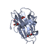
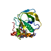
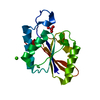
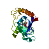
| Entry | Database: PDB / ID: 2zot | ||||||
|---|---|---|---|---|---|---|---|
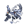
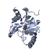
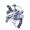
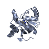
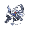
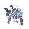
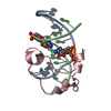
| Title | Crystal structure of human F-spondin reeler domain (fragment 1) | ||||||
 Components Components | Spondin-1 | ||||||
 Keywords Keywords |  CELL ADHESION / beta-sandwich / extracellular protein / Extracellular matrix / Glycoprotein / Secreted CELL ADHESION / beta-sandwich / extracellular protein / Extracellular matrix / Glycoprotein / Secreted | ||||||
| Function / homology |  Function and homology information Function and homology informationpositive regulation of amyloid precursor protein catabolic process / Defective B3GALTL causes PpS / O-glycosylation of TSR domain-containing proteins / positive regulation of protein processing / extracellular matrix structural constituent / LBD domain binding / negative regulation of amyloid-beta formation / extracellular matrix / protein processing / cell adhesion ...positive regulation of amyloid precursor protein catabolic process / Defective B3GALTL causes PpS / O-glycosylation of TSR domain-containing proteins / positive regulation of protein processing / extracellular matrix structural constituent / LBD domain binding / negative regulation of amyloid-beta formation / extracellular matrix / protein processing / cell adhesion / endoplasmic reticulum lumen / extracellular space / metal ion bindingSimilarity search - Function | ||||||
| Biological species |  Homo sapiens (human) Homo sapiens (human) | ||||||
| Method | X-RAY DIFFRACTION / SYNCHROTRON / SAD / Resolution: 2.7 Å | ||||||
 Authors Authors | Nagae, M. / Nogi, T. / Takagi, J. | ||||||
 Citation Citation | Journal: Acta Crystallogr.,Sect.D / Year: 2008 Title: Structure of the F-spondin reeler domain reveals a unique beta-sandwich fold with a deformable disulfide-bonded loop Authors: Nagae, M. / Nishikawa, K. / Yasui, N. / Yamasaki, M. / Nogi, T. / Takagi, J. | ||||||
| History |
|
- Structure visualization
| Structure viewer | Molecule: MolmilJmol/JSmol |
|---|
- Downloads & links
-Download
| PDBx/mmCIF format | 2zot.cif.gz | 113.7 KB | Display | PDBx/mmCIF format |
|---|---|---|---|---|
| PDB format | pdb2zot.ent.gz | 93.9 KB | Display | PDB format |
| PDBx/mmJSON format | 2zot.json.gz | Tree view | PDBx/mmJSON format | |
| Others |  Other downloads Other downloads |
-Validation report
| Arichive directory | https://data.pdbj.org/pub/pdb/validation_reports/zo/2zotftp://data.pdbj.org/pub/pdb/validation_reports/zo/2zot | HTTPS FTP |
|---|
-Related structure data
-Links
 PDBj
PDBj- Assembly
| Deposited unit | 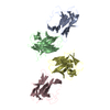
| ||||||||
|---|---|---|---|---|---|---|---|---|---|
| 1 | 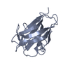
| ||||||||
| 2 | 
| ||||||||
| 3 | 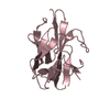
| ||||||||
| 4 | 
| ||||||||
| Unit cell |
|
-Components
| #1: Protein | Mass: 19412.717 Da / Num. of mol.: 4 / Fragment: reeler domain Source method: isolated from a genetically manipulated source Source: (gene. exp.) Homo sapiens (human) / Gene: SPON1, KIAA0762, VSGP / Plasmid: pcDNA3.1/Myc-His(+) vector / Cell line (production host): HEK293T cell / Production host: Homo sapiens (human) / References: UniProt: Q9HCB6 |
|---|
-Experimental details
-Experiment
| Experiment | Method: X-RAY DIFFRACTION / Number of used crystals: 2 |
|---|
- Sample preparation
| Crystal | Density Matthews: 2.06 Å3/Da / Density % sol: 40.2 % |
|---|---|
| Crystal grow | Temperature: 293 K / Method: vapor diffusion, hanging drop / pH: 5.5 Details: 0.2M bis-tris(pH 5.5), 0.4M ammonium acetate, 25% polyethylene glycol(PEG) 3350, VAPOR DIFFUSION, HANGING DROP, temperature 293K |
-Data collection
| Diffraction |
| |||||||||||||||
|---|---|---|---|---|---|---|---|---|---|---|---|---|---|---|---|---|
| Diffraction source |
| |||||||||||||||
| Detector |
| |||||||||||||||
| Radiation |
| |||||||||||||||
| Radiation wavelength |
| |||||||||||||||
| Reflection | Resolution: 2.7→100 Å / Num. all: 17075 / Num. obs: 16670 / % possible obs: 100 % / Redundancy: 4.9 % / Rsym value: 0.083 / Net I/av σ(I): 7.3 / Net I/σ(I): 9 | |||||||||||||||
| Reflection shell | Resolution: 2.7→2.8 Å / Redundancy: 3 % / Mean I/σ(I) obs: 2.038 / Num. unique all: 1724 / Rsym value: 0.488 / % possible all: 99.7 |

- Processing
| Software |
| ||||||||||||||||||||||||||||||||||||||||||||||||||||||||||||||||||||||||||||||||||||||||||||||||||||||||||||||||||||||||||||||||||||||||||||||||||||||||||||||||||||||||||
|---|---|---|---|---|---|---|---|---|---|---|---|---|---|---|---|---|---|---|---|---|---|---|---|---|---|---|---|---|---|---|---|---|---|---|---|---|---|---|---|---|---|---|---|---|---|---|---|---|---|---|---|---|---|---|---|---|---|---|---|---|---|---|---|---|---|---|---|---|---|---|---|---|---|---|---|---|---|---|---|---|---|---|---|---|---|---|---|---|---|---|---|---|---|---|---|---|---|---|---|---|---|---|---|---|---|---|---|---|---|---|---|---|---|---|---|---|---|---|---|---|---|---|---|---|---|---|---|---|---|---|---|---|---|---|---|---|---|---|---|---|---|---|---|---|---|---|---|---|---|---|---|---|---|---|---|---|---|---|---|---|---|---|---|---|---|---|---|---|---|---|---|
| Refinement | Method to determine structure: SAD / Resolution: 2.7→57.54 Å / Cor.coef. Fo:Fc: 0.903 / Cor.coef. Fo:Fc free: 0.835 / SU B: 43.47 / SU ML: 0.398 / TLS residual ADP flag: LIKELY RESIDUAL / Cross valid method: THROUGHOUT / ESU R Free: 0.457 / Stereochemistry target values: MAXIMUM LIKELIHOOD
| ||||||||||||||||||||||||||||||||||||||||||||||||||||||||||||||||||||||||||||||||||||||||||||||||||||||||||||||||||||||||||||||||||||||||||||||||||||||||||||||||||||||||||
| Solvent computation | Ion probe radii: 0.8 Å / Shrinkage radii: 0.8 Å / VDW probe radii: 1.2 Å / Solvent model: MASK | ||||||||||||||||||||||||||||||||||||||||||||||||||||||||||||||||||||||||||||||||||||||||||||||||||||||||||||||||||||||||||||||||||||||||||||||||||||||||||||||||||||||||||
| Displacement parameters | Biso mean: 39.394 Å2
| ||||||||||||||||||||||||||||||||||||||||||||||||||||||||||||||||||||||||||||||||||||||||||||||||||||||||||||||||||||||||||||||||||||||||||||||||||||||||||||||||||||||||||
| Refinement step | Cycle: LAST / Resolution: 2.7→57.54 Å
| ||||||||||||||||||||||||||||||||||||||||||||||||||||||||||||||||||||||||||||||||||||||||||||||||||||||||||||||||||||||||||||||||||||||||||||||||||||||||||||||||||||||||||
| Refine LS restraints |
| ||||||||||||||||||||||||||||||||||||||||||||||||||||||||||||||||||||||||||||||||||||||||||||||||||||||||||||||||||||||||||||||||||||||||||||||||||||||||||||||||||||||||||
| LS refinement shell | Resolution: 2.7→2.77 Å / Total num. of bins used: 20
| ||||||||||||||||||||||||||||||||||||||||||||||||||||||||||||||||||||||||||||||||||||||||||||||||||||||||||||||||||||||||||||||||||||||||||||||||||||||||||||||||||||||||||
| Refinement TLS params. | Method: refined / Refine-ID: X-RAY DIFFRACTION
| ||||||||||||||||||||||||||||||||||||||||||||||||||||||||||||||||||||||||||||||||||||||||||||||||||||||||||||||||||||||||||||||||||||||||||||||||||||||||||||||||||||||||||
| Refinement TLS group |
|
