- PDB-2zmj: Crystal Structure of Rat Vitamin D Receptor Bound to Adamantyl Vi... -
+
Open data
ID or keywords:
Loading...
-
Basic information
Entry
Database: PDB / ID: 2zmj
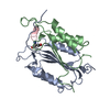
Title
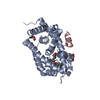
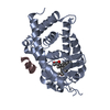
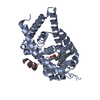
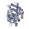
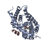
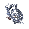
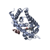
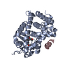
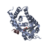
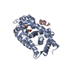
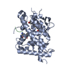
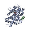
Crystal Structure of Rat Vitamin D Receptor Bound to Adamantyl Vitamin D Analogs: Structural Basis for Vitamin D Receptor Antagonism and/or Partial Agonism
Components
Mediator of RNA polymerase II transcription subunit 1
negative regulation of bone trabecula formation / Vitamin D (calciferol) metabolism / SUMOylation of intracellular receptors / Nuclear Receptor transcription pathway / bile acid nuclear receptor activity / response to bile acid / dense fibrillar component / positive regulation of parathyroid hormone secretion / apoptotic process involved in mammary gland involution / phosphate ion transmembrane transport ...negative regulation of bone trabecula formation / Vitamin D (calciferol) metabolism / SUMOylation of intracellular receptors / Nuclear Receptor transcription pathway / bile acid nuclear receptor activity / response to bile acid / dense fibrillar component / positive regulation of parathyroid hormone secretion / apoptotic process involved in mammary gland involution / phosphate ion transmembrane transport / cellular response to vitamin D / vitamin D binding / calcitriol binding / positive regulation of apoptotic process involved in mammary gland involution / vitamin D response element binding / lithocholic acid binding / mediator complex / positive regulation of keratinocyte differentiation / negative regulation of ossification / positive regulation of vitamin D receptor signaling pathway / vitamin D receptor signaling pathway / bile acid signaling pathway / intestinal absorption / response to aldosterone / mammary gland branching involved in pregnancy / regulation of calcium ion transport / decidualization / negative regulation of keratinocyte proliferation / heterochromatin / nuclear retinoid X receptor binding / T-tubule / lactation / skeletal system development / apoptotic signaling pathway / transcription coregulator activity / animal organ morphogenesis / euchromatin / mRNA transcription by RNA polymerase II / cell morphogenesis / nuclear matrix / response to calcium ion / intracellular calcium ion homeostasis / RNA polymerase II transcription regulator complex / cellular response to amyloid-beta / calcium ion transport / nuclear receptor activity / response to estradiol / heart development / sequence-specific DNA binding / cell differentiation / receptor complex / RNA polymerase II cis-regulatory region sequence-specific DNA binding / DNA-binding transcription factor activity / negative regulation of cell population proliferation / negative regulation of DNA-templated transcription / regulation of DNA-templated transcription / positive regulation of gene expression / regulation of transcription by RNA polymerase II / perinuclear region of cytoplasm / negative regulation of transcription by RNA polymerase II / positive regulation of transcription by RNA polymerase II / DNA binding / zinc ion binding / nucleus Similarity search - Function
Mediator complex, subunit Med1 / Mediator of RNA polymerase II transcription subunit 1 / Vitamin D receptor / VDR, DNA-binding domain / Retinoid X Receptor / Retinoid X Receptor / Nuclear hormone receptor / Nuclear hormones receptors DNA-binding region signature. / Zinc finger, nuclear hormone receptor-type / Zinc finger, C4 type (two domains) ...Mediator complex, subunit Med1 / Mediator of RNA polymerase II transcription subunit 1 / Vitamin D receptor / VDR, DNA-binding domain / Retinoid X Receptor / Retinoid X Receptor / Nuclear hormone receptor / Nuclear hormones receptors DNA-binding region signature. / Zinc finger, nuclear hormone receptor-type / Zinc finger, C4 type (two domains) / Nuclear hormone receptors DNA-binding domain profile. / c4 zinc finger in nuclear hormone receptors / Nuclear hormone receptor, ligand-binding domain / Nuclear hormone receptor-like domain superfamily / Ligand-binding domain of nuclear hormone receptor / Nuclear receptor (NR) ligand-binding (LBD) domain profile. / Ligand binding domain of hormone receptors / Zinc finger, NHR/GATA-type / Orthogonal Bundle / Mainly Alpha Similarity search - Domain/homology
Chem-MI4 / Mediator of RNA polymerase II transcription subunit 1 / Vitamin D3 receptor Similarity search - Component
In the structure databanks used in Yorodumi, some data are registered as the other names, "COVID-19 virus" and "2019-nCoV". Here are the details of the virus and the list of structure data.
Jan 31, 2019. EMDB accession codes are about to change! (news from PDBe EMDB page)
EMDB accession codes are about to change! (news from PDBe EMDB page)
The allocation of 4 digits for EMDB accession codes will soon come to an end. Whilst these codes will remain in use, new EMDB accession codes will include an additional digit and will expand incrementally as the available range of codes is exhausted. The current 4-digit format prefixed with “EMD-” (i.e. EMD-XXXX) will advance to a 5-digit format (i.e. EMD-XXXXX), and so on. It is currently estimated that the 4-digit codes will be depleted around Spring 2019, at which point the 5-digit format will come into force.
The EM Navigator/Yorodumi systems omit the EMD- prefix.
Related info.:Q: What is EMD? / ID/Accession-code notation in Yorodumi/EM Navigator
Yorodumi is a browser for structure data from EMDB, PDB, SASBDB, etc.
This page is also the successor to EM Navigator detail page, and also detail information page/front-end page for Omokage search.
The word "yorodu" (or yorozu) is an old Japanese word meaning "ten thousand". "mi" (miru) is to see.
Related info.:EMDB / PDB / SASBDB / Comparison of 3 databanks / Yorodumi Search / Aug 31, 2016. New EM Navigator & Yorodumi / Yorodumi Papers / Jmol/JSmol / Function and homology information / Changes in new EM Navigator and Yorodumi
 Movie
Movie Controller

 Yorodumi
 Open data
 Basic information
 Components
 Keywords
 TRANSCRIPTION / NUCLEAR RECEPTOR-ANTAGONIST COMPLEX / DNA-binding / Metal-binding /
  Function and homology information
 Authors
 Citation
 Structure visualization
 Downloads & links
 Other downloads


 PDBj
PDBj


 Assembly


 Mass: 534.812 Da / Num. of mol.: 1 / Source method: obtained synthetically / Formula: C36H54O3
 Mass: 18.015 Da / Num. of mol.: 16 / Source method: isolated from a natural source / Formula: H2O
 Sample preparation
 / Beamline: AR-NW12A / Wavelength: 1 Å
 Processing