 Movie
Movie Controller

[English] 日本語
 Yorodumi
- PDB-2zhy: Crystal structure of a pduO-type ATP:cobalamin adenosyltransferas... -
+ Open data

- Basic information
| Entry | Database: PDB / ID: 2zhy | ||||||
|---|---|---|---|---|---|---|---|
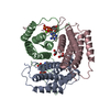
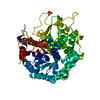
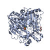
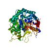
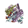
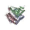
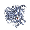
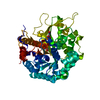
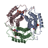
| Title | Crystal structure of a pduO-type ATP:cobalamin adenosyltransferase from Burkholderia thailandensis | ||||||
 Components Components | ATP:cob(I)alamin adenosyltransferase, putative | ||||||
 Keywords Keywords |  TRANSFERASE / helix bundle TRANSFERASE / helix bundle | ||||||
| Function / homology |  Function and homology information Function and homology informationcorrinoid adenosyltransferase activity / Transferases; Transferring alkyl or aryl groups, other than methyl groups / cobalamin biosynthetic process / ATP binding / metal ion bindingSimilarity search - Function | ||||||
| Biological species |  Burkholderia thailandensis (bacteria) Burkholderia thailandensis (bacteria) | ||||||
| Method | X-RAY DIFFRACTION / SYNCHROTRON / Resolution: 1.8 Å | ||||||
 Authors Authors | Moon, J.H. / Park, A.K. / Jang, E.H. / Kim, H.S. / Chi, Y.M. | ||||||
 Citation Citation | Journal: Proteins / Year: 2008 Title: Crystal structure of a PduO-type ATP:cobalamin adenosyltransferase from Burkholderia thailandensis. Authors: Moon, J.H. / Park, A.K. / Jang, E.H. / Kim, H.S. / Chi, Y.M. | ||||||
| History |
|
- Structure visualization
| Structure viewer | Molecule: MolmilJmol/JSmol |
|---|
- Downloads & links
-Download
| PDBx/mmCIF format | 2zhy.cif.gz | 102.8 KB | Display | PDBx/mmCIF format |
|---|---|---|---|---|
| PDB format | pdb2zhy.ent.gz | 79.5 KB | Display | PDB format |
| PDBx/mmJSON format | 2zhy.json.gz | Tree view | PDBx/mmJSON format | |
| Others |  Other downloads Other downloads |
-Validation report
| Arichive directory | https://data.pdbj.org/pub/pdb/validation_reports/zh/2zhyftp://data.pdbj.org/pub/pdb/validation_reports/zh/2zhy | HTTPS FTP |
|---|
-Related structure data


-Links
 PDBj
PDBj- Assembly
| Deposited unit | 
| ||||||||
|---|---|---|---|---|---|---|---|---|---|
| 1 |
| ||||||||
| Unit cell |
|
-Components
| #1: Protein | Mass: 19522.150 Da / Num. of mol.: 3 Source method: isolated from a genetically manipulated source Source: (gene. exp.) Burkholderia thailandensis (bacteria) / Gene: pduO / Plasmid: pET-28a / Production host: Escherichia coli (E. coli) / Strain (production host): BL21(DE3) / References: UniProt: Q2SZ09, corrinoid adenosyltransferase#2: Water | ChemComp-HOH / | Water Mass: 18.015 Da / Num. of mol.: 200 / Source method: isolated from a natural source / Formula: H2O Mass: 18.015 Da / Num. of mol.: 200 / Source method: isolated from a natural source / Formula: H2O |
|---|
-Experimental details
-Experiment
| Experiment | Method: X-RAY DIFFRACTION / Number of used crystals: 1 |
|---|
- Sample preparation
| Crystal | Density Matthews: 2.63 Å3/Da / Density % sol: 53.29 % |
|---|---|
| Crystal grow | Temperature: 295 K / Method: vapor diffusion, hanging drop / pH: 5.7 Details: 0.1M Na Citrate, 22% Isopropanol, 12% PEG 4000, pH 5.7, VAPOR DIFFUSION, HANGING DROP, temperature 295K |
-Data collection
| Diffraction | Mean temperature: 100 K |
|---|---|
| Diffraction source | Source: SYNCHROTRON / Site: PAL/PLS  / Beamline: 4A / Wavelength: 0.9999 Å / Beamline: 4A / Wavelength: 0.9999 Å |
| Detector | Type: ADSC QUANTUM 210 / Detector: CCD / Date: Dec 7, 2007 / Details: mirrors |
| Radiation | Monochromator: double crystals / Protocol: SINGLE WAVELENGTH / Monochromatic (M) / Laue (L): M / Scattering type: x-ray |
| Radiation wavelength | Wavelength: 0.9999 Å / Relative weight: 1 |
| Reflection | Resolution: 1.8→50 Å / Num. all: 54263 / Num. obs: 54261 / % possible obs: 94.2 % / Observed criterion σ(F): 0 / Redundancy: 7.6 % / Biso Wilson estimate: 14.7 Å2 / Limit h max: 29 / Limit h min: 0 / Limit k max: 82 / Limit k min: 0 / Limit l max: 86 / Limit l min: 0 / Observed criterion F max: 54211.48 / Observed criterion F min: 0.32 / Rmerge(I) obs: 0.091 / Χ2: 1.713 / Net I/σ(I): 10.3 |
| Reflection shell | Resolution: 1.8→1.86 Å / Redundancy: 2.3 % / Rmerge(I) obs: 0.344 / Num. unique all: 3908 / Χ2: 0.524 / % possible all: 68.9 |
- Processing
| Software |
| ||||||||||||||||||||||||||||||||||||||||||||||||||||||||||||||||||||||||||||||||||||||||||
|---|---|---|---|---|---|---|---|---|---|---|---|---|---|---|---|---|---|---|---|---|---|---|---|---|---|---|---|---|---|---|---|---|---|---|---|---|---|---|---|---|---|---|---|---|---|---|---|---|---|---|---|---|---|---|---|---|---|---|---|---|---|---|---|---|---|---|---|---|---|---|---|---|---|---|---|---|---|---|---|---|---|---|---|---|---|---|---|---|---|---|---|
| Refinement | Resolution: 1.8→47.3 Å / Rfactor Rfree error: 0.005 / Occupancy max: 1 / Occupancy min: 1 / Cross valid method: THROUGHOUT / σ(F): 0 / Stereochemistry target values: Engh & Huber
| ||||||||||||||||||||||||||||||||||||||||||||||||||||||||||||||||||||||||||||||||||||||||||
| Solvent computation | Solvent model: CNS bulk solvent model used / Bsol: 64.305 Å2 / ksol: 0.4 e/Å3 | ||||||||||||||||||||||||||||||||||||||||||||||||||||||||||||||||||||||||||||||||||||||||||
| Displacement parameters | Biso max: 67.45 Å2 / Biso mean: 28.473 Å2 / Biso min: 10.56 Å2
| ||||||||||||||||||||||||||||||||||||||||||||||||||||||||||||||||||||||||||||||||||||||||||
| Refine analyze |
| ||||||||||||||||||||||||||||||||||||||||||||||||||||||||||||||||||||||||||||||||||||||||||
| Refinement step | Cycle: LAST / Resolution: 1.8→47.3 Å
| ||||||||||||||||||||||||||||||||||||||||||||||||||||||||||||||||||||||||||||||||||||||||||
| Refine LS restraints |
| ||||||||||||||||||||||||||||||||||||||||||||||||||||||||||||||||||||||||||||||||||||||||||
| LS refinement shell | Refine-ID: X-RAY DIFFRACTION / Total num. of bins used: 8
| ||||||||||||||||||||||||||||||||||||||||||||||||||||||||||||||||||||||||||||||||||||||||||
| Xplor file |
|
