 Movie
Movie Controller


+ Open data

- Basic information
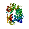
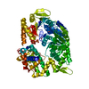
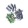
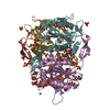
| Entry | Database: PDB / ID: 2z41 | ||||||
|---|---|---|---|---|---|---|---|
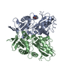
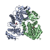
| Title | Crystal Structure Analysis of the Ski2-type RNA helicase | ||||||
 Components Components | putative ski2-type helicase | ||||||
 Keywords Keywords |  HYDROLASE / RNA helicase / DNA helicase HYDROLASE / RNA helicase / DNA helicase | ||||||
| Biological species |   Pyrococcus horikoshii (archaea) Pyrococcus horikoshii (archaea) | ||||||
| Method | X-RAY DIFFRACTION / SYNCHROTRON / SAD / Resolution: 3.51 Å | ||||||
 Authors Authors | Nakashima, T. / Zhang, X. / Kakuta, Y. / Yao, M. / Tanaka, I. / Kimura, M. | ||||||
 Citation Citation | Journal: Protein Sci. / Year: 2008 Title: Crystal structure of an archaeal Ski2p-like protein from Pyrococcus horikoshii OT3 Authors: Zhang, X. / Nakashima, T. / Kakuta, Y. / Yao, M. / Tanaka, I. / Kimura, M. | ||||||
| History |
|
- Structure visualization
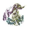
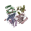
| Structure viewer | Molecule:  MolmilJmol/JSmol MolmilJmol/JSmol |
|---|
- Downloads & links
-Download
| PDBx/mmCIF format | 2z41.cif.gz | 30 KB | Display | PDBx/mmCIF format |
|---|---|---|---|---|
| PDB format | pdb2z41.ent.gz | 18.7 KB | Display | PDB format |
| PDBx/mmJSON format | 2z41.json.gz | Tree view | PDBx/mmJSON format | |
| Others |  Other downloads Other downloads |
-Validation report
| Arichive directory | https://data.pdbj.org/pub/pdb/validation_reports/z4/2z41ftp://data.pdbj.org/pub/pdb/validation_reports/z4/2z41 | HTTPS FTP |
|---|
-Related structure data
| Similar structure data |
|---|
-Links
 PDBj
PDBj
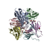
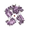
- Assembly
| Deposited unit | 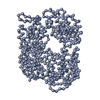
| ||||||||
|---|---|---|---|---|---|---|---|---|---|
| 1 |
| ||||||||
| Unit cell |
| ||||||||
| Details | The biological unit is not determined. It may be a monomer. |
-Components
| #1: Protein | Mass: 55166.117 Da / Num. of mol.: 1 Source method: isolated from a genetically manipulated source Source: (gene. exp.) Pyrococcus horikoshii (archaea) / Strain: OT3 / Gene: PH1280 / Plasmid: pET-22b / Species (production host): Escherichia coli / Production host:  Escherichia coli BL21(DE3) (bacteria) / Strain (production host): BL21(DE3) Escherichia coli BL21(DE3) (bacteria) / Strain (production host): BL21(DE3)References: Hydrolases; Acting on acid anhydrides; In phosphorus-containing anhydrides |
|---|---|
| #2: Chemical | ChemComp-MG /   Mass: 24.305 Da / Num. of mol.: 1 / Source method: obtained synthetically / Formula: Mg Mass: 24.305 Da / Num. of mol.: 1 / Source method: obtained synthetically / Formula: Mg |
| Sequence details | BECAUSE THE ELECTRON DENSITY WAS POOR, THE SIDE CHAINS WERE PARTIALLY FITTED BY A HOMOLOGY MODEL ...BECAUSE THE ELECTRON DENSITY WAS POOR, THE SIDE CHAINS WERE PARTIALLY FITTED BY A HOMOLOGY MODEL MADE FROM RECQ PROTEIN (PDB ID: 1OYW). ALTHOUGH REFINEMENT |
-Experimental details
-Experiment
| Experiment | Method: X-RAY DIFFRACTION / Number of used crystals: 1 |
|---|
- Sample preparation
| Crystal | Density Matthews: 3.75 Å3/Da / Density % sol: 67.24 % |
|---|---|
| Crystal grow | Temperature: 293 K / Method: vapor diffusion, hanging drop / pH: 4.2 Details: 10% 2-propanol, 0.2M Lithium sulfate, 0.1M phosphate-citrate buffer, pH 4.2, VAPOR DIFFUSION, HANGING DROP, temperature 293.0K |
-Data collection
| Diffraction | Mean temperature: 100 K |
|---|---|
| Diffraction source | Source: SYNCHROTRON / Site: SPring-8  / Beamline: BL41XU / Wavelength: 0.97904 Å / Beamline: BL41XU / Wavelength: 0.97904 Å |
| Detector | Type: ADSC QUANTUM 315 / Detector: CCD / Date: Apr 9, 2007 |
| Radiation | Protocol: SINGLE WAVELENGTH / Monochromatic (M) / Laue (L): M / Scattering type: x-ray |
| Radiation wavelength | Wavelength: 0.97904 Å / Relative weight: 1 |
| Reflection | Resolution: 3.51→50 Å / Num. all: 16159 / Num. obs: 16159 / % possible obs: 99.9 % / Observed criterion σ(F): 0 / Observed criterion σ(I): 0 / Redundancy: 6.3 % / Biso Wilson estimate: 53.5 Å2 / Rmerge(I) obs: 0.129 / Rsym value: 0.137 |
| Reflection shell | Resolution: 3.51→3.63 Å / Redundancy: 5.4 % / Rmerge(I) obs: 0.454 / Num. unique all: 2969 / Rsym value: 0.474 / % possible all: 99.9 |
- Processing
| Software |
| |||||||||||||||||||||||||||||||||||||||||||||||||
|---|---|---|---|---|---|---|---|---|---|---|---|---|---|---|---|---|---|---|---|---|---|---|---|---|---|---|---|---|---|---|---|---|---|---|---|---|---|---|---|---|---|---|---|---|---|---|---|---|---|---|
| Refinement | Method to determine structure: SAD / Resolution: 3.51→20 Å / Cross valid method: THROUGHOUT / σ(F): 0 / Stereochemistry target values: Engh & Huber Details: The R factor and free-R factor were calculated for the coordinates containing the side chains.
| |||||||||||||||||||||||||||||||||||||||||||||||||
| Refinement step | Cycle: LAST / Resolution: 3.51→20 Å
| |||||||||||||||||||||||||||||||||||||||||||||||||
| Refine LS restraints |
| |||||||||||||||||||||||||||||||||||||||||||||||||
| LS refinement shell |
|
