 Movie
Movie Controller


+ Open data

- Basic information
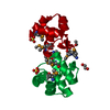
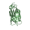
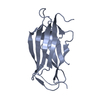
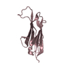
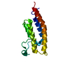
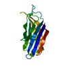
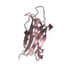
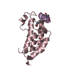
| Entry | Database: PDB / ID: 2z0u | ||||||
|---|---|---|---|---|---|---|---|
| Title | Crystal structure of C2 domain of KIBRA protein | ||||||
 Components Components | WW domain-containing protein 1 | ||||||
 Keywords Keywords | LIPID BINDING PROTEIN /  C2 domain / Alternative splicing / Coiled coil / Cytoplasm / Phosphorylation / Polymorphism / Structural Genomics / NPPSFA / National Project on Protein Structural and Functional Analyses / RIKEN Structural Genomics/Proteomics Initiative / RSGI C2 domain / Alternative splicing / Coiled coil / Cytoplasm / Phosphorylation / Polymorphism / Structural Genomics / NPPSFA / National Project on Protein Structural and Functional Analyses / RIKEN Structural Genomics/Proteomics Initiative / RSGI | ||||||
| Function / homology |  Function and homology informationregulation of intracellular transport / regulation of hippo signaling / negative regulation of organ growth / Signaling by Hippo / NOTCH3 Intracellular Domain Regulates Transcription / establishment of cell polarity / negative regulation of hippo signaling / ruffle membrane / kinase binding / cell migration ...regulation of intracellular transport / regulation of hippo signaling / negative regulation of organ growth / Signaling by Hippo / NOTCH3 Intracellular Domain Regulates Transcription / establishment of cell polarity / negative regulation of hippo signaling / ruffle membrane / kinase binding / cell migration / positive regulation of MAPK cascade / transcription coactivator activity / molecular adaptor activity / negative regulation of cell population proliferation / regulation of DNA-templated transcription / perinuclear region of cytoplasm / negative regulation of transcription by RNA polymerase II / protein-containing complex / nucleus / cytosol / cytoplasm Function and homology informationregulation of intracellular transport / regulation of hippo signaling / negative regulation of organ growth / Signaling by Hippo / NOTCH3 Intracellular Domain Regulates Transcription / establishment of cell polarity / negative regulation of hippo signaling / ruffle membrane / kinase binding / cell migration ...regulation of intracellular transport / regulation of hippo signaling / negative regulation of organ growth / Signaling by Hippo / NOTCH3 Intracellular Domain Regulates Transcription / establishment of cell polarity / negative regulation of hippo signaling / ruffle membrane / kinase binding / cell migration / positive regulation of MAPK cascade / transcription coactivator activity / molecular adaptor activity / negative regulation of cell population proliferation / regulation of DNA-templated transcription / perinuclear region of cytoplasm / negative regulation of transcription by RNA polymerase II / protein-containing complex / nucleus / cytosol / cytoplasmSimilarity search - Function | ||||||
| Biological species |  Homo sapiens (human) Homo sapiens (human) | ||||||
| Method | X-RAY DIFFRACTION / SYNCHROTRON / SAD / Resolution: 2.2 Å | ||||||
 Authors Authors | Murayama, K. / Kato-Murayama, M. / Terada, T. / Shirouzu, M. / Yokoyama, S. / RIKEN Structural Genomics/Proteomics Initiative (RSGI) | ||||||
 Citation Citation | Journal: To be Published Title: Crystal structure of C2 domain of KIBRA protein Authors: Murayama, K. / Kato-Murayama, M. / Terada, T. / Shirouzu, M. / Yokoyama, S. | ||||||
| History |
|
- Structure visualization
| Structure viewer | Molecule: MolmilJmol/JSmol |
|---|
- Downloads & links
-Download
| PDBx/mmCIF format | 2z0u.cif.gz | 60.7 KB | Display | PDBx/mmCIF format |
|---|---|---|---|---|
| PDB format | pdb2z0u.ent.gz | 48.6 KB | Display | PDB format |
| PDBx/mmJSON format | 2z0u.json.gz | Tree view | PDBx/mmJSON format | |
| Others |  Other downloads Other downloads |
-Validation report
| Arichive directory | https://data.pdbj.org/pub/pdb/validation_reports/z0/2z0uftp://data.pdbj.org/pub/pdb/validation_reports/z0/2z0u | HTTPS FTP |
|---|
-Related structure data
| Similar structure data | |
|---|---|
| Other databases |


-Links
 PDBj
PDBj


- Assembly
| Deposited unit | 
| ||||||||
|---|---|---|---|---|---|---|---|---|---|
| 1 | 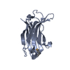
| ||||||||
| 2 | 
| ||||||||
| 3 |
| ||||||||
| Unit cell |
|
-Components
| #1: Protein | Mass: 17094.055 Da / Num. of mol.: 2 / Fragment: C2 domain Source method: isolated from a genetically manipulated source Source: (gene. exp.) Homo sapiens (human) / Plasmid: PK060110-34 / Production host: cell free protein synthesis (others) / References: UniProt: Q8IX03#2: Water | ChemComp-HOH / | Water Mass: 18.015 Da / Num. of mol.: 118 / Source method: isolated from a natural source / Formula: H2O Mass: 18.015 Da / Num. of mol.: 118 / Source method: isolated from a natural source / Formula: H2O |
|---|
-Experimental details
-Experiment
| Experiment | Method: X-RAY DIFFRACTION / Number of used crystals: 1 |
|---|
- Sample preparation
| Crystal | Density Matthews: 2.11 Å3/Da / Density % sol: 41.73 % |
|---|---|
| Crystal grow | Temperature: 293 K / Method: vapor diffusion, hanging drop / Details: VAPOR DIFFUSION, HANGING DROP, temperature 293K |
-Data collection
| Diffraction | Mean temperature: 100 K |
|---|---|
| Diffraction source | Source: SYNCHROTRON / Site: SPring-8  / Beamline: BL26B2 / Wavelength: 0.979 Å / Beamline: BL26B2 / Wavelength: 0.979 Å |
| Detector | Type: RIGAKU JUPITER 210 / Detector: CCD / Date: Mar 5, 2007 |
| Radiation | Protocol: SINGLE WAVELENGTH / Monochromatic (M) / Laue (L): M / Scattering type: x-ray |
| Radiation wavelength | Wavelength: 0.979 Å / Relative weight: 1 |
| Reflection | Resolution: 2.2→44.46 Å / Num. obs: 15280 / % possible obs: 99.8 % / Observed criterion σ(F): -3 / Redundancy: 7 % / Biso Wilson estimate: 17.8 Å2 / Rsym value: 0.074 / Net I/σ(I): 22.6 |
| Reflection shell | Resolution: 2.2→2.28 Å / Rsym value: 0.303 / % possible all: 99.9 |
- Processing
| Software |
| ||||||||||||||||||||||||||||||||||||||||||||||||||||||||||||||||||||||||||||||||
|---|---|---|---|---|---|---|---|---|---|---|---|---|---|---|---|---|---|---|---|---|---|---|---|---|---|---|---|---|---|---|---|---|---|---|---|---|---|---|---|---|---|---|---|---|---|---|---|---|---|---|---|---|---|---|---|---|---|---|---|---|---|---|---|---|---|---|---|---|---|---|---|---|---|---|---|---|---|---|---|---|---|
| Refinement | Method to determine structure: SAD / Resolution: 2.2→44.46 Å / Rfactor Rfree error: 0.007 / Data cutoff high absF: 1255189.7 / Data cutoff low absF: 0 / Isotropic thermal model: RESTRAINED / Cross valid method: THROUGHOUT / σ(F): 0
| ||||||||||||||||||||||||||||||||||||||||||||||||||||||||||||||||||||||||||||||||
| Solvent computation | Solvent model: FLAT MODEL / Bsol: 31.8635 Å2 / ksol: 0.364328 e/Å3 | ||||||||||||||||||||||||||||||||||||||||||||||||||||||||||||||||||||||||||||||||
| Displacement parameters | Biso mean: 33.8 Å2
| ||||||||||||||||||||||||||||||||||||||||||||||||||||||||||||||||||||||||||||||||
| Refine analyze |
| ||||||||||||||||||||||||||||||||||||||||||||||||||||||||||||||||||||||||||||||||
| Refinement step | Cycle: LAST / Resolution: 2.2→44.46 Å
| ||||||||||||||||||||||||||||||||||||||||||||||||||||||||||||||||||||||||||||||||
| Refine LS restraints |
| ||||||||||||||||||||||||||||||||||||||||||||||||||||||||||||||||||||||||||||||||
| LS refinement shell | Resolution: 2.2→2.34 Å / Rfactor Rfree error: 0.019 / Total num. of bins used: 6
| ||||||||||||||||||||||||||||||||||||||||||||||||||||||||||||||||||||||||||||||||
| Xplor file |
|
