 Movie
Movie Controller

[English] 日本語
 Yorodumi
- PDB-2yw6: Structural studies of N terminal deletion mutant of Dps from Myco... -
+ Open data

- Basic information
| Entry | Database: PDB / ID: 2yw6 | ||||||
|---|---|---|---|---|---|---|---|
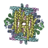
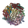
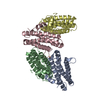
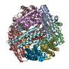
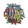
| Title | Structural studies of N terminal deletion mutant of Dps from Mycobacterium smegmatis | ||||||
 Components Components | DNA protection during starvation protein | ||||||
 Keywords Keywords |  OXIDOREDUCTASE / DNA-BINDING PROTEIN / Quarternary assembly / Ferroxidation OXIDOREDUCTASE / DNA-BINDING PROTEIN / Quarternary assembly / Ferroxidation | ||||||
| Function / homology |  Function and homology informationOxidoreductases; Oxidizing metal ions / oxidoreductase activity, acting on metal ions / nucleoid / ferric iron binding / intracellular iron ion homeostasis / DNA binding / cytoplasm Function and homology informationOxidoreductases; Oxidizing metal ions / oxidoreductase activity, acting on metal ions / nucleoid / ferric iron binding / intracellular iron ion homeostasis / DNA binding / cytoplasmSimilarity search - Function | ||||||
| Biological species |  Mycobacterium smegmatis (bacteria) Mycobacterium smegmatis (bacteria) | ||||||
| Method | X-RAY DIFFRACTION / MOLECULAR REPLACEMENT / Resolution: 2.53 Å | ||||||
 Authors Authors | Roy, S. / Saraswathi, R. / Gupta, S. / Sekar, K. / Chatterji, D. / Vijayan, M. | ||||||
 Citation Citation | Journal: J.Mol.Biol. / Year: 2007 Title: Role of N and C-terminal Tails in DNA Binding and Assembly in Dps: Structural Studies of Mycobacterium smegmatis Dps Deletion Mutants Authors: Roy, S. / Saraswathi, R. / Gupta, S. / Sekar, K. / Chatterji, D. / Vijayan, M. | ||||||
| History |
|
- Structure visualization
| Structure viewer | Molecule: MolmilJmol/JSmol |
|---|
- Downloads & links
-Download
| PDBx/mmCIF format | 2yw6.cif.gz | 102.6 KB | Display | PDBx/mmCIF format |
|---|---|---|---|---|
| PDB format | pdb2yw6.ent.gz | 79.6 KB | Display | PDB format |
| PDBx/mmJSON format | 2yw6.json.gz | Tree view | PDBx/mmJSON format | |
| Others |  Other downloads Other downloads |
-Validation report
| Arichive directory | https://data.pdbj.org/pub/pdb/validation_reports/yw/2yw6ftp://data.pdbj.org/pub/pdb/validation_reports/yw/2yw6 | HTTPS FTP |
|---|
-Related structure data
| Related structure data |  2yw7C  1veiS S: Starting model for refinement C: citing same article ( |
|---|---|
| Similar structure data |

-Links
 PDBj
PDBj


- Assembly
| Deposited unit | 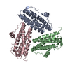
| ||||||||
|---|---|---|---|---|---|---|---|---|---|
| 1 | 
| ||||||||
| Unit cell |
|
-Components
| #1: Protein | Mass: 20298.809 Da / Num. of mol.: 3 Source method: isolated from a genetically manipulated source Source: (gene. exp.) Mycobacterium smegmatis (bacteria) / Gene: dps / Plasmid: pET-msdpsdelta9 / Species (production host): Escherichia coli / Production host: Escherichia coli BL21(DE3) (bacteria) / Strain (production host): BL21DE3References: UniProt: Q8VP75, UniProt: P0C558*PLUS, Oxidoreductases; Oxidizing metal ions#2: Water | ChemComp-HOH / | Water Mass: 18.015 Da / Num. of mol.: 185 / Source method: isolated from a natural source / Formula: H2O Mass: 18.015 Da / Num. of mol.: 185 / Source method: isolated from a natural source / Formula: H2O |
|---|
-Experimental details
-Experiment
| Experiment | Method: X-RAY DIFFRACTION / Number of used crystals: 1 |
|---|
- Sample preparation
| Crystal | Density Matthews: 1.93 Å3/Da / Density % sol: 36.12 % |
|---|---|
| Crystal grow | Temperature: 293 K / Method: microbath under oil / pH: 8 Details: protein buffer in 50mM Tris-HCl (pH8.0), 0.2M Calcium chloride, 0.1M HEPES-Na pH7.5, 28% polyethelene glycol 400, Microbath under oil, temperature 293K |
-Data collection
| Diffraction | Mean temperature: 100 K |
|---|---|
| Diffraction source | Source: ROTATING ANODE / Type: RIGAKU RU200 / Wavelength: 1.5418 Å |
| Detector | Type: MAR scanner 300 mm plate / Detector: IMAGE PLATE / Date: Oct 22, 2005 |
| Radiation | Monochromator: OSMIC MIRROR / Protocol: SINGLE WAVELENGTH / Monochromatic (M) / Laue (L): M / Scattering type: x-ray |
| Radiation wavelength | Wavelength: 1.5418 Å / Relative weight: 1 |
| Reflection | Resolution: 2.53→30 Å / Num. obs: 15936 / % possible obs: 94.4 % / Observed criterion σ(F): 0 / Redundancy: 17 % / Biso Wilson estimate: 45.1 Å2 / Rmerge(I) obs: 0.098 / Net I/σ(I): 28 |
| Reflection shell | Resolution: 2.53→2.69 Å / Rmerge(I) obs: 0.497 / Mean I/σ(I) obs: 3.83 / Num. unique all: 1411 / % possible all: 88.3 |
- Processing
| Software |
| ||||||||||||||||||||||||||||||||||||
|---|---|---|---|---|---|---|---|---|---|---|---|---|---|---|---|---|---|---|---|---|---|---|---|---|---|---|---|---|---|---|---|---|---|---|---|---|---|
| Refinement | Method to determine structure: MOLECULAR REPLACEMENT Starting model: 1VEI Resolution: 2.53→28.62 Å / Rfactor Rfree error: 0.009 / Data cutoff high absF: 2414985.54 / Data cutoff low absF: 0 / Isotropic thermal model: RESTRAINED / Cross valid method: THROUGHOUT / σ(F): 0 / Stereochemistry target values: Engh & Huber
| ||||||||||||||||||||||||||||||||||||
| Solvent computation | Solvent model: FLAT MODEL / Bsol: 46.6917 Å2 / ksol: 0.374647 e/Å3 | ||||||||||||||||||||||||||||||||||||
| Displacement parameters | Biso mean: 46.6 Å2
| ||||||||||||||||||||||||||||||||||||
| Refine analyze |
| ||||||||||||||||||||||||||||||||||||
| Refinement step | Cycle: LAST / Resolution: 2.53→28.62 Å
| ||||||||||||||||||||||||||||||||||||
| Refine LS restraints |
| ||||||||||||||||||||||||||||||||||||
| LS refinement shell | Resolution: 2.53→2.69 Å / Rfactor Rfree error: 0.03 / Total num. of bins used: 6
| ||||||||||||||||||||||||||||||||||||
| Xplor file |
|
