 Movie
Movie Controller

[English] 日本語
 Yorodumi
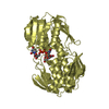
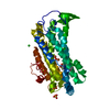
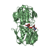
- PDB-2yvw: Crystal structure of UDP-N-acetylglucosamine 1-carboxyvinyltransf... -
+ Open data

- Basic information
| Entry | Database: PDB / ID: 2yvw | ||||||
|---|---|---|---|---|---|---|---|
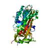
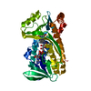
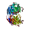
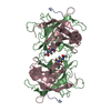
| Title | Crystal structure of UDP-N-acetylglucosamine 1-carboxyvinyltransferase from Aquifex aeolicus VF5 | ||||||
 Components Components | UDP-N-acetylglucosamine 1-carboxyvinyltransferase | ||||||
 Keywords Keywords | TRANSFERASE / PEPTIDOGLYCAN / UDP-N-ACETYLGLUCOSAMINE / Structural Genomics / NPPSFA / National Project on Protein Structural and Functional Analyses / RIKEN Structural Genomics/Proteomics Initiative / RSGI | ||||||
| Function / homology |  Function and homology informationUDP-N-acetylglucosamine 1-carboxyvinyltransferase / UDP-N-acetylglucosamine 1-carboxyvinyltransferase activity / UDP-N-acetylgalactosamine biosynthetic process / peptidoglycan biosynthetic process / cell wall organization / regulation of cell shape / cell cycle / cell division / cytoplasm Function and homology informationUDP-N-acetylglucosamine 1-carboxyvinyltransferase / UDP-N-acetylglucosamine 1-carboxyvinyltransferase activity / UDP-N-acetylgalactosamine biosynthetic process / peptidoglycan biosynthetic process / cell wall organization / regulation of cell shape / cell cycle / cell division / cytoplasmSimilarity search - Function | ||||||
| Biological species |   Aquifex aeolicus (bacteria) Aquifex aeolicus (bacteria) | ||||||
| Method | X-RAY DIFFRACTION / SYNCHROTRON / MOLECULAR REPLACEMENT / Resolution: 1.81 Å | ||||||
 Authors Authors | Kitamuta, Y. / Yokoyama, S. / Kuramitsu, S. / RIKEN Structural Genomics/Proteomics Initiative (RSGI) | ||||||
 Citation Citation | Journal: To be Published Title: Crystal structure of UDP-N-acetylglucosamine 1-carboxyvinyltransferase from Aquifex aeolicus VF5 Authors: Kitamura, Y. / Yokoyama, S. / Kuramitsu, S. | ||||||
| History |
|
- Structure visualization
| Structure viewer | Molecule: MolmilJmol/JSmol |
|---|
- Downloads & links
-Download
| PDBx/mmCIF format | 2yvw.cif.gz | 101.2 KB | Display | PDBx/mmCIF format |
|---|---|---|---|---|
| PDB format | pdb2yvw.ent.gz | 76.4 KB | Display | PDB format |
| PDBx/mmJSON format | 2yvw.json.gz | Tree view | PDBx/mmJSON format | |
| Others |  Other downloads Other downloads |
-Validation report
| Arichive directory | https://data.pdbj.org/pub/pdb/validation_reports/yv/2yvwftp://data.pdbj.org/pub/pdb/validation_reports/yv/2yvw | HTTPS FTP |
|---|
-Related structure data
| Related structure data | 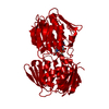 1uaeS S: Starting model for refinement |
|---|---|
| Similar structure data | |
| Other databases |


-Links
 PDBj
PDBj
- Assembly
| Deposited unit | 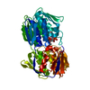
| ||||||||
|---|---|---|---|---|---|---|---|---|---|
| 1 |
| ||||||||
| Unit cell |
|
-Components
| #1: Protein | / Enoylpyruvate transferase / UDP-N-acetylglucosamine enolpyruvyl transferase / EPT Mass: 47322.496 Da / Num. of mol.: 1 Source method: isolated from a genetically manipulated source Source: (gene. exp.) Aquifex aeolicus (bacteria) / Strain: VF5 / Plasmid: pET-21a / Production host: Escherichia coli (E. coli)References: UniProt: O67315, UDP-N-acetylglucosamine 1-carboxyvinyltransferase |
|---|---|
| #2: Chemical | ChemComp-PO4 / Phosphate  Mass: 94.971 Da / Num. of mol.: 1 / Source method: obtained synthetically / Formula: PO4 Mass: 94.971 Da / Num. of mol.: 1 / Source method: obtained synthetically / Formula: PO4 |
| #3: Chemical | ChemComp-EPU / 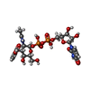  Mass: 677.400 Da / Num. of mol.: 1 / Source method: obtained synthetically / Formula: C20H29N3O19P2 Mass: 677.400 Da / Num. of mol.: 1 / Source method: obtained synthetically / Formula: C20H29N3O19P2 |
| #4: Water | ChemComp-HOH / Water Mass: 18.015 Da / Num. of mol.: 284 / Source method: isolated from a natural source / Formula: H2O Mass: 18.015 Da / Num. of mol.: 284 / Source method: isolated from a natural source / Formula: H2O |
-Experimental details
-Experiment
| Experiment | Method: X-RAY DIFFRACTION / Number of used crystals: 1 |
|---|
- Sample preparation
| Crystal | Density Matthews: 3.09 Å3/Da / Density % sol: 60.2 % |
|---|---|
| Crystal grow | Temperature: 291 K / Method: microbatch / pH: 7.4 Details: 40% (v/v) PEG 400, 0.1M Imidazole, pH 7.4, MICRO BATCH, temperature 291K |
-Data collection
| Diffraction | Mean temperature: 100 K |
|---|---|
| Diffraction source | Source: SYNCHROTRON / Site: SPring-8  / Beamline: BL26B1 / Wavelength: 1 Å / Beamline: BL26B1 / Wavelength: 1 Å |
| Detector | Type: RIGAKU JUPITER 210 / Detector: CCD / Date: Mar 21, 2007 |
| Radiation | Monochromator: Fixed exit Si double crystal monochromator / Protocol: SINGLE WAVELENGTH / Monochromatic (M) / Laue (L): M / Scattering type: x-ray |
| Radiation wavelength | Wavelength: 1 Å / Relative weight: 1 |
| Reflection | Resolution: 1.81→50 Å / Num. obs: 52283 / % possible obs: 98.7 % / Redundancy: 10.4 % / Biso Wilson estimate: 14.2 Å2 / Rmerge(I) obs: 0.067 / Net I/σ(I): 33.47 |
| Reflection shell | Resolution: 1.81→1.87 Å / Redundancy: 8.9 % / Rmerge(I) obs: 0.329 / Mean I/σ(I) obs: 6.24 / % possible all: 90.8 |
- Processing
| Software |
| ||||||||||||||||||||||||||||||||||||||||||||||||||||||||||||||||||||||||||||||||
|---|---|---|---|---|---|---|---|---|---|---|---|---|---|---|---|---|---|---|---|---|---|---|---|---|---|---|---|---|---|---|---|---|---|---|---|---|---|---|---|---|---|---|---|---|---|---|---|---|---|---|---|---|---|---|---|---|---|---|---|---|---|---|---|---|---|---|---|---|---|---|---|---|---|---|---|---|---|---|---|---|---|
| Refinement | Method to determine structure: MOLECULAR REPLACEMENT Starting model: PDB ENTRY 1UAE Resolution: 1.81→43.64 Å / Rfactor Rfree error: 0.004 / Data cutoff high absF: 91872.23 / Data cutoff low absF: 0 / Isotropic thermal model: RESTRAINED / Cross valid method: THROUGHOUT / σ(F): 0 / Stereochemistry target values: MAXIMUM LIKELIHOOD
| ||||||||||||||||||||||||||||||||||||||||||||||||||||||||||||||||||||||||||||||||
| Solvent computation | Solvent model: FLAT MODEL / Bsol: 46.8358 Å2 / ksol: 0.366653 e/Å3 | ||||||||||||||||||||||||||||||||||||||||||||||||||||||||||||||||||||||||||||||||
| Displacement parameters | Biso mean: 23.6 Å2
| ||||||||||||||||||||||||||||||||||||||||||||||||||||||||||||||||||||||||||||||||
| Refine analyze |
| ||||||||||||||||||||||||||||||||||||||||||||||||||||||||||||||||||||||||||||||||
| Refinement step | Cycle: LAST / Resolution: 1.81→43.64 Å
| ||||||||||||||||||||||||||||||||||||||||||||||||||||||||||||||||||||||||||||||||
| Refine LS restraints |
| ||||||||||||||||||||||||||||||||||||||||||||||||||||||||||||||||||||||||||||||||
| LS refinement shell | Resolution: 1.81→1.92 Å / Rfactor Rfree error: 0.013 / Total num. of bins used: 6
| ||||||||||||||||||||||||||||||||||||||||||||||||||||||||||||||||||||||||||||||||
| Xplor file |
|
