 Movie
Movie Controller

[English] 日本語
 Yorodumi
- PDB-2yr6: Crystal structure of L-phenylalanine oxidase from Psuedomonas sp.P501 -
+ Open data

- Basic information
| Entry | Database: PDB / ID: 2yr6 | |||||||||
|---|---|---|---|---|---|---|---|---|---|---|
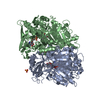
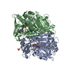
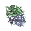
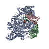
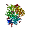
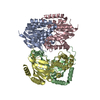
| Title | Crystal structure of L-phenylalanine oxidase from Psuedomonas sp.P501 | |||||||||
 Components Components | Pro-enzyme of L-phenylalanine oxidase | |||||||||
 Keywords Keywords |  OXIDOREDUCTASE / L-phenylalanine oxidase / amino oxidase / flavoenzyme OXIDOREDUCTASE / L-phenylalanine oxidase / amino oxidase / flavoenzyme | |||||||||
| Function / homology |  Function and homology informationphenylalanine 2-monooxygenase / phenylalanine 2-monooxygenase activity / nucleotide binding Function and homology informationphenylalanine 2-monooxygenase / phenylalanine 2-monooxygenase activity / nucleotide bindingSimilarity search - Function | |||||||||
| Biological species |  Pseudomonas sp. P-501 (bacteria) Pseudomonas sp. P-501 (bacteria) | |||||||||
| Method | X-RAY DIFFRACTION / SYNCHROTRON / MOLECULAR REPLACEMENT / Resolution: 1.35 Å | |||||||||
 Authors Authors | Ida, K. / Kurabayashi, M. / Suguro, M. / Suzuki, H. | |||||||||
 Citation Citation | Journal: J.Biol.Chem. / Year: 2008 Title: Structural basis of proteolytic activation of L-phenylalanine oxidase from Pseudomonas sp. P-501. Authors: Ida, K. / Kurabayashi, M. / Suguro, M. / Hiruma, Y. / Hikima, T. / Yamomoto, M. / Suzuki, H. | |||||||||
| History |
|
- Structure visualization
| Structure viewer | Molecule: MolmilJmol/JSmol |
|---|
- Downloads & links
-Download
| PDBx/mmCIF format | 2yr6.cif.gz | 621.6 KB | Display | PDBx/mmCIF format |
|---|---|---|---|---|
| PDB format | pdb2yr6.ent.gz | 504.3 KB | Display | PDB format |
| PDBx/mmJSON format | 2yr6.json.gz | Tree view | PDBx/mmJSON format | |
| Others |  Other downloads Other downloads |
-Validation report
| Arichive directory | https://data.pdbj.org/pub/pdb/validation_reports/yr/2yr6ftp://data.pdbj.org/pub/pdb/validation_reports/yr/2yr6 | HTTPS FTP |
|---|
-Related structure data
| Related structure data |  2yr4SC  2yr5C  2yr7 2yr8 2yr9 S: Starting model for refinement C: citing same article ( |
|---|---|
| Similar structure data |


-Links
 PDBj
PDBj- Assembly
| Deposited unit | 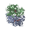
| ||||||||
|---|---|---|---|---|---|---|---|---|---|
| 1 |
| ||||||||
| Unit cell |
|
-Components
-Protein , 1 types, 2 molecules AB
| #1: Protein | Mass: 77899.664 Da / Num. of mol.: 2 Source method: isolated from a genetically manipulated source Source: (gene. exp.) Pseudomonas sp. P-501 (bacteria) / Plasmid: pET22b / Species (production host): Escherichia coli / Production host: Escherichia coli BL21(DE3) (bacteria) / Strain (production host): BL21(DE3) / References: UniProt: Q5W9R9, phenylalanine 2-monooxygenase |
|---|
-Non-polymers , 5 types, 1965 molecules 


| #2: Chemical | Sulfate Mass: 96.063 Da / Num. of mol.: 2 / Source method: obtained synthetically / Formula: SO4 Mass: 96.063 Da / Num. of mol.: 2 / Source method: obtained synthetically / Formula: SO4#3: Chemical | Flavin adenine dinucleotide Mass: 785.550 Da / Num. of mol.: 2 / Source method: obtained synthetically / Formula: C27H33N9O15P2 / Comment: FAD*YM Mass: 785.550 Da / Num. of mol.: 2 / Source method: obtained synthetically / Formula: C27H33N9O15P2 / Comment: FAD*YM#4: Chemical | Anthranilic acid Type: L-peptide linking / Mass: 137.136 Da / Num. of mol.: 2 / Source method: obtained synthetically / Formula: C7H7NO2 Type: L-peptide linking / Mass: 137.136 Da / Num. of mol.: 2 / Source method: obtained synthetically / Formula: C7H7NO2#5: Chemical | Glycerol Mass: 92.094 Da / Num. of mol.: 2 / Source method: obtained synthetically / Formula: C3H8O3 Mass: 92.094 Da / Num. of mol.: 2 / Source method: obtained synthetically / Formula: C3H8O3#6: Water | ChemComp-HOH / | WaterMass: 18.015 Da / Num. of mol.: 1957 / Source method: isolated from a natural source / Formula: H2O |
|---|
-Experimental details
-Experiment
| Experiment | Method: X-RAY DIFFRACTION / Number of used crystals: 1 |
|---|
- Sample preparation
| Crystal | Density Matthews: 2.5 Å3/Da / Density % sol: 50.71 % |
|---|---|
| Crystal grow | Temperature: 293 K / Method: vapor diffusion / pH: 7.5 Details: 0.1M HEPES pH7.5, 1.0M ammonium sulfate, VAPOR DIFFUSION, temperature 293K |
-Data collection
| Diffraction | Mean temperature: 100 K |
|---|---|
| Diffraction source | Source: SYNCHROTRON / Site: Photon Factory  / Beamline: AR-NW12A / Wavelength: 1 Å / Beamline: AR-NW12A / Wavelength: 1 Å |
| Detector | Type: ADSC QUANTUM 210 / Detector: CCD / Date: Nov 3, 2005 |
| Radiation | Protocol: SINGLE WAVELENGTH / Monochromatic (M) / Laue (L): M / Scattering type: x-ray |
| Radiation wavelength | Wavelength: 1 Å / Relative weight: 1 |
| Reflection | Resolution: 1.35→34.18 Å / Num. obs: 339311 / % possible obs: 99.9 % / Redundancy: 5.5 % / Rmerge(I) obs: 0.089 |
| Reflection shell | Resolution: 1.35→1.42 Å / Redundancy: 5.3 % / Rmerge(I) obs: 0.342 / % possible all: 100 |
- Processing
| Software |
| |||||||||||||||||||||||||||||||||||||||||||||||||||||||||||||||||||||||||||||||||||||||||||||||||||||||||||||||||||||||||||||||||||||||||||||||||
|---|---|---|---|---|---|---|---|---|---|---|---|---|---|---|---|---|---|---|---|---|---|---|---|---|---|---|---|---|---|---|---|---|---|---|---|---|---|---|---|---|---|---|---|---|---|---|---|---|---|---|---|---|---|---|---|---|---|---|---|---|---|---|---|---|---|---|---|---|---|---|---|---|---|---|---|---|---|---|---|---|---|---|---|---|---|---|---|---|---|---|---|---|---|---|---|---|---|---|---|---|---|---|---|---|---|---|---|---|---|---|---|---|---|---|---|---|---|---|---|---|---|---|---|---|---|---|---|---|---|---|---|---|---|---|---|---|---|---|---|---|---|---|---|---|---|---|
| Refinement | Method to determine structure: MOLECULAR REPLACEMENT Starting model: 2YR4 Resolution: 1.35→34.18 Å / Cor.coef. Fo:Fc: 0.984 / Cor.coef. Fo:Fc free: 0.975 / SU B: 1.122 / SU ML: 0.021 / Cross valid method: THROUGHOUT / ESU R: 0.035 / ESU R Free: 0.037 / Stereochemistry target values: MAXIMUM LIKELIHOOD / Details: HYDROGENS HAVE BEEN ADDED IN THE RIDING POSITIONS
| |||||||||||||||||||||||||||||||||||||||||||||||||||||||||||||||||||||||||||||||||||||||||||||||||||||||||||||||||||||||||||||||||||||||||||||||||
| Solvent computation | Ion probe radii: 0.8 Å / Shrinkage radii: 0.8 Å / VDW probe radii: 1.2 Å / Solvent model: BABINET MODEL WITH MASK | |||||||||||||||||||||||||||||||||||||||||||||||||||||||||||||||||||||||||||||||||||||||||||||||||||||||||||||||||||||||||||||||||||||||||||||||||
| Displacement parameters | Biso mean: 12.85 Å2
| |||||||||||||||||||||||||||||||||||||||||||||||||||||||||||||||||||||||||||||||||||||||||||||||||||||||||||||||||||||||||||||||||||||||||||||||||
| Refinement step | Cycle: LAST / Resolution: 1.35→34.18 Å
| |||||||||||||||||||||||||||||||||||||||||||||||||||||||||||||||||||||||||||||||||||||||||||||||||||||||||||||||||||||||||||||||||||||||||||||||||
| Refine LS restraints |
| |||||||||||||||||||||||||||||||||||||||||||||||||||||||||||||||||||||||||||||||||||||||||||||||||||||||||||||||||||||||||||||||||||||||||||||||||
| LS refinement shell | Resolution: 1.35→1.385 Å / Total num. of bins used: 20
|
