 Movie
Movie Controller


+ Open data

- Basic information
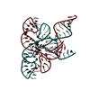
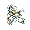
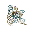
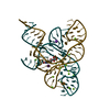
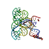
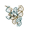
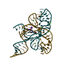
| Entry | Database: PDB / ID: 2yie | ||||||
|---|---|---|---|---|---|---|---|
| Title | Crystal structure of a F. nucleatum FMN riboswitch bound to FMN | ||||||
 Components Components | (FMN RIBOSWITCH ) x 2 ) x 2 | ||||||
 Keywords Keywords | RNA | ||||||
| Function / homology | FLAVIN MONONUCLEOTIDE / : / : / RNA / RNA (> 10) Function and homology information Function and homology information | ||||||
| Biological species |  FUSOBACTERIUM NUCLEATUM SUBSP. NUCLEATUM (bacteria) FUSOBACTERIUM NUCLEATUM SUBSP. NUCLEATUM (bacteria) | ||||||
| Method | X-RAY DIFFRACTION / SYNCHROTRON / MOLECULAR REPLACEMENT / Resolution: 2.941 Å | ||||||
 Authors Authors | Vicens, Q. / Mondragon, E. / Batey, R.T. | ||||||
 Citation Citation | Journal: Nucleic Acids Res. / Year: 2011 Title: Molecular Sensing by the Aptamer Domain of the Fmn Riboswitch: A General Model for Ligand Binding by Conformational Selection Authors: Vicens, Q. / Mondragon, E. / Batey, R.T. | ||||||
| History |
|
- Structure visualization
| Structure viewer | Molecule: MolmilJmol/JSmol |
|---|
- Downloads & links
-Download
| PDBx/mmCIF format | 2yie.cif.gz | 138.9 KB | Display | PDBx/mmCIF format |
|---|---|---|---|---|
| PDB format | pdb2yie.ent.gz | 109.4 KB | Display | PDB format |
| PDBx/mmJSON format | 2yie.json.gz | Tree view | PDBx/mmJSON format | |
| Others |  Other downloads Other downloads |
-Validation report
| Arichive directory | https://data.pdbj.org/pub/pdb/validation_reports/yi/2yieftp://data.pdbj.org/pub/pdb/validation_reports/yi/2yie | HTTPS FTP |
|---|
-Related structure data
| Related structure data |  2yifC 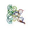 3f2qS C: citing same article ( S: Starting model for refinement |
|---|---|
| Similar structure data |


-Links
 PDBj
PDBj


- Assembly
| Deposited unit | 
| ||||||||
|---|---|---|---|---|---|---|---|---|---|
| 1 |
| ||||||||
| Unit cell |
|
-Components
-RNA chain , 2 types, 2 molecules XZ
| #1: RNA chain | Mass: 17575.324 Da / Num. of mol.: 1 / Fragment: APTAMER DOMAIN, RESIDUES 2495-2544 / Mutation: YES / Source method: obtained synthetically / Details: APTAMER DOMAIN BOUND TO FLAVIN MONONUCLEOTIDE Source: (synth.) FUSOBACTERIUM NUCLEATUM SUBSP. NUCLEATUM (bacteria)References: GenBank: 20095250 |
|---|---|
| #2: RNA chain | Mass: 18661.000 Da / Num. of mol.: 1 / Fragment: APTAMER DOMAIN, RESIDUES 2550-2601 / Mutation: YES / Source method: obtained synthetically / Details: APTAMER DOMAIN BOUND TO FLAVIN MONONUCLEOTIDE Source: (synth.) FUSOBACTERIUM NUCLEATUM SUBSP. NUCLEATUM (bacteria)References: GenBank: 20095250 |
-Non-polymers , 4 types, 46 molecules 


| #3: Chemical | ChemComp-MG /  Mass: 24.305 Da / Num. of mol.: 14 / Source method: obtained synthetically / Formula: Mg Mass: 24.305 Da / Num. of mol.: 14 / Source method: obtained synthetically / Formula: Mg#4: Chemical |  Mass: 39.098 Da / Num. of mol.: 2 / Source method: obtained synthetically / Formula: K Mass: 39.098 Da / Num. of mol.: 2 / Source method: obtained synthetically / Formula: K#5: Chemical | ChemComp-FMN / | Flavin mononucleotide Mass: 456.344 Da / Num. of mol.: 1 / Source method: obtained synthetically / Formula: C17H21N4O9P Mass: 456.344 Da / Num. of mol.: 1 / Source method: obtained synthetically / Formula: C17H21N4O9P#6: Water | ChemComp-HOH / | WaterMass: 18.015 Da / Num. of mol.: 29 / Source method: isolated from a natural source / Formula: H2O |
|---|
-Details
| Compound details | ENGINEERED RESIDUE IN CHAIN X, A 2542 TO U ENGINEERED RESIDUE IN CHAIN X, G 2543 TO C ENGINEERED ...ENGINEERED |
|---|
-Experimental details
-Experiment
| Experiment | Method: X-RAY DIFFRACTION / Number of used crystals: 1 |
|---|
- Sample preparation
| Crystal | Density Matthews: 1.5 Å3/Da / Density % sol: 64 % Description: DATA WAS COLLECTED BY ANNIE HEROUX AS PART OF THE MACROMOLECULAR CRYSTALLOGRAPHY RESEARCH RESOURCE AT BROOKHAVEN NSLS |
|---|---|
| Crystal grow | Details: RNA WAS CRYSTALLIZED FROM 15% PEG-MONOMETHYL 550 ETHER, 0.1 M MGCL2, 0.05 M NA-HEPES PH 7.0, 1 MM FMN |
-Data collection
| Diffraction | Mean temperature: 100 K |
|---|---|
| Diffraction source | Source: SYNCHROTRON / Site: NSLS  / Beamline: X25 / Wavelength: 0.9795 / Beamline: X25 / Wavelength: 0.9795 |
| Detector | Type: ADSC CCD / Detector: CCD / Date: Aug 3, 2009 |
| Radiation | Protocol: SINGLE WAVELENGTH / Monochromatic (M) / Laue (L): M / Scattering type: x-ray |
| Radiation wavelength | Wavelength: 0.9795 Å / Relative weight: 1 |
| Reflection | Resolution: 2.95→40 Å / Num. obs: 17834 / % possible obs: 99.3 % / Observed criterion σ(I): 2 / Redundancy: 4.7 % / Biso Wilson estimate: 80.36 Å2 / Rmerge(I) obs: 0.05 / Net I/σ(I): 21.7 |
| Reflection shell | Resolution: 2.95→3.06 Å / Redundancy: 3.4 % / Rmerge(I) obs: 0.48 / Mean I/σ(I) obs: 3.4 / % possible all: 100 |
- Processing
| Software |
| |||||||||||||||||||||||||||||||||||||||||||||||||||||||||||||||||||||||||||||||||||||||||||||||||||||||||||||||||||||||||||||||||||||||||||||||||||||||||||||||||||||||||||||||||||||||||||||||||||||||||||||||||||||||||||||||||||||||||||||||||||||||||||||||||||||||||||||||||||||||||||||||||||||||||||||||||||||||||||||||||||||
|---|---|---|---|---|---|---|---|---|---|---|---|---|---|---|---|---|---|---|---|---|---|---|---|---|---|---|---|---|---|---|---|---|---|---|---|---|---|---|---|---|---|---|---|---|---|---|---|---|---|---|---|---|---|---|---|---|---|---|---|---|---|---|---|---|---|---|---|---|---|---|---|---|---|---|---|---|---|---|---|---|---|---|---|---|---|---|---|---|---|---|---|---|---|---|---|---|---|---|---|---|---|---|---|---|---|---|---|---|---|---|---|---|---|---|---|---|---|---|---|---|---|---|---|---|---|---|---|---|---|---|---|---|---|---|---|---|---|---|---|---|---|---|---|---|---|---|---|---|---|---|---|---|---|---|---|---|---|---|---|---|---|---|---|---|---|---|---|---|---|---|---|---|---|---|---|---|---|---|---|---|---|---|---|---|---|---|---|---|---|---|---|---|---|---|---|---|---|---|---|---|---|---|---|---|---|---|---|---|---|---|---|---|---|---|---|---|---|---|---|---|---|---|---|---|---|---|---|---|---|---|---|---|---|---|---|---|---|---|---|---|---|---|---|---|---|---|---|---|---|---|---|---|---|---|---|---|---|---|---|---|---|---|---|---|---|---|---|---|---|---|---|---|---|---|---|---|---|---|---|---|---|---|---|---|---|---|---|---|---|---|---|---|---|---|---|---|---|---|---|---|---|---|---|---|---|---|---|---|---|---|---|---|---|---|---|---|---|---|---|---|---|---|---|---|---|---|
| Refinement | Method to determine structure: MOLECULAR REPLACEMENT Starting model: PDB ENTRY 3F2Q Resolution: 2.941→26.292 Å / SU ML: 0.35 / σ(F): 0 / Phase error: 24.5 / Stereochemistry target values: ML Details: THE GAMMA PHOSPHATE OF GTP IS DISORDERED ON CHAINS X AND Z
| |||||||||||||||||||||||||||||||||||||||||||||||||||||||||||||||||||||||||||||||||||||||||||||||||||||||||||||||||||||||||||||||||||||||||||||||||||||||||||||||||||||||||||||||||||||||||||||||||||||||||||||||||||||||||||||||||||||||||||||||||||||||||||||||||||||||||||||||||||||||||||||||||||||||||||||||||||||||||||||||||||||
| Solvent computation | Shrinkage radii: 0.83 Å / VDW probe radii: 1.1 Å / Solvent model: FLAT BULK SOLVENT MODEL / Bsol: 43.744 Å2 / ksol: 0.247 e/Å3 | |||||||||||||||||||||||||||||||||||||||||||||||||||||||||||||||||||||||||||||||||||||||||||||||||||||||||||||||||||||||||||||||||||||||||||||||||||||||||||||||||||||||||||||||||||||||||||||||||||||||||||||||||||||||||||||||||||||||||||||||||||||||||||||||||||||||||||||||||||||||||||||||||||||||||||||||||||||||||||||||||||||
| Displacement parameters | Biso mean: 96.2 Å2
| |||||||||||||||||||||||||||||||||||||||||||||||||||||||||||||||||||||||||||||||||||||||||||||||||||||||||||||||||||||||||||||||||||||||||||||||||||||||||||||||||||||||||||||||||||||||||||||||||||||||||||||||||||||||||||||||||||||||||||||||||||||||||||||||||||||||||||||||||||||||||||||||||||||||||||||||||||||||||||||||||||||
| Refinement step | Cycle: LAST / Resolution: 2.941→26.292 Å
| |||||||||||||||||||||||||||||||||||||||||||||||||||||||||||||||||||||||||||||||||||||||||||||||||||||||||||||||||||||||||||||||||||||||||||||||||||||||||||||||||||||||||||||||||||||||||||||||||||||||||||||||||||||||||||||||||||||||||||||||||||||||||||||||||||||||||||||||||||||||||||||||||||||||||||||||||||||||||||||||||||||
| Refine LS restraints |
| |||||||||||||||||||||||||||||||||||||||||||||||||||||||||||||||||||||||||||||||||||||||||||||||||||||||||||||||||||||||||||||||||||||||||||||||||||||||||||||||||||||||||||||||||||||||||||||||||||||||||||||||||||||||||||||||||||||||||||||||||||||||||||||||||||||||||||||||||||||||||||||||||||||||||||||||||||||||||||||||||||||
| LS refinement shell |
| |||||||||||||||||||||||||||||||||||||||||||||||||||||||||||||||||||||||||||||||||||||||||||||||||||||||||||||||||||||||||||||||||||||||||||||||||||||||||||||||||||||||||||||||||||||||||||||||||||||||||||||||||||||||||||||||||||||||||||||||||||||||||||||||||||||||||||||||||||||||||||||||||||||||||||||||||||||||||||||||||||||
| Refinement TLS params. | Method: refined / Refine-ID: X-RAY DIFFRACTION
| |||||||||||||||||||||||||||||||||||||||||||||||||||||||||||||||||||||||||||||||||||||||||||||||||||||||||||||||||||||||||||||||||||||||||||||||||||||||||||||||||||||||||||||||||||||||||||||||||||||||||||||||||||||||||||||||||||||||||||||||||||||||||||||||||||||||||||||||||||||||||||||||||||||||||||||||||||||||||||||||||||||
| Refinement TLS group |
|
