 Movie
Movie Controller


+ Open data

- Basic information
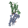
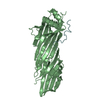
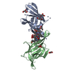
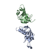
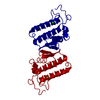
| Entry | Database: PDB / ID: 2yhc | ||||||
|---|---|---|---|---|---|---|---|
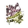
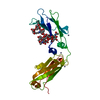
| Title | Structure of BamD from E. coli | ||||||
 Components Components | UPF0169 LIPOPROTEIN YFIO | ||||||
 Keywords Keywords |  MEMBRANE PROTEIN / LIPOPROTEIN / ESSENTIAL BAM COMPONENT MEMBRANE PROTEIN / LIPOPROTEIN / ESSENTIAL BAM COMPONENT | ||||||
| Function / homology |  Function and homology information Function and homology informationBam protein complex / Gram-negative-bacterium-type cell outer membrane assembly / protein insertion into membrane / cell outer membrane / membraneSimilarity search - Function | ||||||
| Biological species |  ESCHERICHIA COLI (E. coli) ESCHERICHIA COLI (E. coli) | ||||||
| Method | X-RAY DIFFRACTION / SYNCHROTRON / MIRAS / Resolution: 1.8 Å | ||||||
 Authors Authors | Zeth, K. / Albrecht, R. | ||||||
 Citation Citation | Journal: J.Biol.Chem. / Year: 2011 Title: Structural Basis of Outer Membrane Protein Biogenesis in Bacteria. Authors: Albrecht, R. / Zeth, K. | ||||||
| History |
|
- Structure visualization
| Structure viewer | Molecule: MolmilJmol/JSmol |
|---|
- Downloads & links
-Download
| PDBx/mmCIF format | 2yhc.cif.gz | 99.5 KB | Display | PDBx/mmCIF format |
|---|---|---|---|---|
| PDB format | pdb2yhc.ent.gz | 77.5 KB | Display | PDB format |
| PDBx/mmJSON format | 2yhc.json.gz | Tree view | PDBx/mmJSON format | |
| Others |  Other downloads Other downloads |
-Validation report
| Arichive directory | https://data.pdbj.org/pub/pdb/validation_reports/yh/2yhcftp://data.pdbj.org/pub/pdb/validation_reports/yh/2yhc | HTTPS FTP |
|---|
-Related structure data


-Links
 PDBj
PDBj


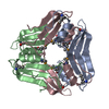
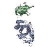
- Assembly
| Deposited unit | 
| ||||||||
|---|---|---|---|---|---|---|---|---|---|
| 1 |
| ||||||||
| Unit cell |
|
-Components
| #1: Protein | Mass: 25959.949 Da / Num. of mol.: 1 / Fragment: RESIDUES 29-245 Source method: isolated from a genetically manipulated source Source: (gene. exp.) ESCHERICHIA COLI (E. coli) / Plasmid: PET24D / Production host: ESCHERICHIA COLI (E. coli) / Strain (production host): BL21(DE3) / References: UniProt: P0AC02 |
|---|---|
| #2: Chemical | ChemComp-URE / Urea  Mass: 60.055 Da / Num. of mol.: 1 / Source method: obtained synthetically / Formula: CH4N2O Mass: 60.055 Da / Num. of mol.: 1 / Source method: obtained synthetically / Formula: CH4N2O |
| #3: Water | ChemComp-HOH / Water Mass: 18.015 Da / Num. of mol.: 84 / Source method: isolated from a natural source / Formula: H2O Mass: 18.015 Da / Num. of mol.: 84 / Source method: isolated from a natural source / Formula: H2O |
-Experimental details
-Experiment
| Experiment | Method: X-RAY DIFFRACTION / Number of used crystals: 1 |
|---|
- Sample preparation
| Crystal | Density Matthews: 1.84 Å3/Da / Density % sol: 33.14 % / Description: NONE |
|---|---|
| Crystal grow | pH: 7 / Details: 30% PEG 4000, 10% I-PROH, 0.1 M HEPES PH 7.5 . |
-Data collection
| Diffraction | Mean temperature: 100 K |
|---|---|
| Diffraction source | Source: SYNCHROTRON / Site: SLS  / Beamline: X10SA / Wavelength: 1 / Beamline: X10SA / Wavelength: 1 |
| Detector | Type: DECTRIS PILATUS 6M / Detector: PIXEL |
| Radiation | Protocol: SINGLE WAVELENGTH / Monochromatic (M) / Laue (L): M / Scattering type: x-ray |
| Radiation wavelength | Wavelength: 1 Å / Relative weight: 1 |
| Reflection | Resolution: 1.8→40 Å / Num. obs: 16896 / % possible obs: 99.4 % / Observed criterion σ(I): 3.9 / Redundancy: 6.2 % / Rmerge(I) obs: 0.04 / Net I/σ(I): 20.3 |
| Reflection shell | Resolution: 1.8→1.9 Å / Rmerge(I) obs: 0.51 / Mean I/σ(I) obs: 3.9 / % possible all: 97.8 |
- Processing
| Software |
| ||||||||||||||||||||||||||||||||||||||||||||||||||||||||||||||||||||||||||||||||||||||||||||||||||||||||||||||||||||||||||||||||||||||||||||||||||||||||||||||||||||||||||||||||||||||
|---|---|---|---|---|---|---|---|---|---|---|---|---|---|---|---|---|---|---|---|---|---|---|---|---|---|---|---|---|---|---|---|---|---|---|---|---|---|---|---|---|---|---|---|---|---|---|---|---|---|---|---|---|---|---|---|---|---|---|---|---|---|---|---|---|---|---|---|---|---|---|---|---|---|---|---|---|---|---|---|---|---|---|---|---|---|---|---|---|---|---|---|---|---|---|---|---|---|---|---|---|---|---|---|---|---|---|---|---|---|---|---|---|---|---|---|---|---|---|---|---|---|---|---|---|---|---|---|---|---|---|---|---|---|---|---|---|---|---|---|---|---|---|---|---|---|---|---|---|---|---|---|---|---|---|---|---|---|---|---|---|---|---|---|---|---|---|---|---|---|---|---|---|---|---|---|---|---|---|---|---|---|---|---|
| Refinement | Method to determine structure: MIRAS Starting model: NONE Resolution: 1.8→28.39 Å / Cor.coef. Fo:Fc: 0.959 / Cor.coef. Fo:Fc free: 0.946 / SU B: 7.041 / SU ML: 0.1 / Cross valid method: THROUGHOUT / ESU R: 0.157 / ESU R Free: 0.139 / Stereochemistry target values: MAXIMUM LIKELIHOOD / Details: HYDROGENS HAVE BEEN ADDED IN THE RIDING POSITIONS.
| ||||||||||||||||||||||||||||||||||||||||||||||||||||||||||||||||||||||||||||||||||||||||||||||||||||||||||||||||||||||||||||||||||||||||||||||||||||||||||||||||||||||||||||||||||||||
| Solvent computation | Ion probe radii: 0.8 Å / Shrinkage radii: 0.8 Å / VDW probe radii: 1.4 Å / Solvent model: MASK | ||||||||||||||||||||||||||||||||||||||||||||||||||||||||||||||||||||||||||||||||||||||||||||||||||||||||||||||||||||||||||||||||||||||||||||||||||||||||||||||||||||||||||||||||||||||
| Displacement parameters | Biso mean: 44.667 Å2
| ||||||||||||||||||||||||||||||||||||||||||||||||||||||||||||||||||||||||||||||||||||||||||||||||||||||||||||||||||||||||||||||||||||||||||||||||||||||||||||||||||||||||||||||||||||||
| Refinement step | Cycle: LAST / Resolution: 1.8→28.39 Å
| ||||||||||||||||||||||||||||||||||||||||||||||||||||||||||||||||||||||||||||||||||||||||||||||||||||||||||||||||||||||||||||||||||||||||||||||||||||||||||||||||||||||||||||||||||||||
| Refine LS restraints |
|