 Movie
Movie Controller


+ Open data

- Basic information
| Entry | Database: PDB / ID: 2yha | ||||||
|---|---|---|---|---|---|---|---|
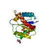
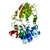
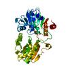
| Title | Crystal Structure of the N. crassa QDE-2 AGO MID-PIWI Domains | ||||||
 Components Components | POST-TRANSCRIPTIONAL GENE SILENCING PROTEIN QDE-2 RNA interference RNA interference | ||||||
 Keywords Keywords | RNA BINDING PROTEIN / ARGONAUTE / MIRNA / SIRNA | ||||||
| Function / homology |  Function and homology information Function and homology information | ||||||
| Biological species |  NEUROSPORA CRASSA (fungus) NEUROSPORA CRASSA (fungus) | ||||||
| Method | X-RAY DIFFRACTION / SYNCHROTRON / MOLECULAR REPLACEMENT / Resolution: 1.85 Å | ||||||
 Authors Authors | Boland, A. / Weichenrieder, O. | ||||||
 Citation Citation | Journal: Proc.Natl.Acad.Sci.USA / Year: 2011 Title: Crystal Structure of the Mid-Piwi Lobe of a Eukaryotic Argonaute Protein Authors: Boland, A. / Huntzinger, E. / Schmidt, S. / Izaurralde, E. / Weichenriede, O. | ||||||
| History |
| ||||||
| Remark 650 | HELIX DETERMINATION METHOD: AUTHOR PROVIDED. |
- Structure visualization
| Structure viewer | Molecule: MolmilJmol/JSmol |
|---|
- Downloads & links
-Download
| PDBx/mmCIF format | 2yha.cif.gz | 150.4 KB | Display | PDBx/mmCIF format |
|---|---|---|---|---|
| PDB format | pdb2yha.ent.gz | 119.1 KB | Display | PDB format |
| PDBx/mmJSON format | 2yha.json.gz | Tree view | PDBx/mmJSON format | |
| Others |  Other downloads Other downloads |
-Validation report
| Arichive directory | https://data.pdbj.org/pub/pdb/validation_reports/yh/2yhaftp://data.pdbj.org/pub/pdb/validation_reports/yh/2yha | HTTPS FTP |
|---|
-Related structure data
| Related structure data | 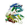 2yhbSC S: Starting model for refinement C: citing same article ( |
|---|---|
| Similar structure data |


-Links
 PDBj
PDBj
- Assembly
| Deposited unit | 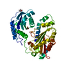
| ||||||||
|---|---|---|---|---|---|---|---|---|---|
| 1 |
| ||||||||
| Unit cell |
|
-Components
| #1: Protein | RNA interference Mass: 42986.453 Da / Num. of mol.: 1 / Fragment: MID-PIWI DOMAINS, RESIDUES 506-786 AND 839-938 Source method: isolated from a genetically manipulated source Details: RESIDUES 787-838 REPLACED BY THREE-RESIDUE LINKER / Source: (gene. exp.) NEUROSPORA CRASSA (fungus) / Plasmid: PETM60 / Production host:  ESCHERICHIA COLI (E. coli) / Strain (production host): K-12 / References: UniProt: Q9P8T1 ESCHERICHIA COLI (E. coli) / Strain (production host): K-12 / References: UniProt: Q9P8T1 |
|---|---|
| #2: Chemical | ChemComp-SO4 / Sulfate  Mass: 96.063 Da / Num. of mol.: 1 / Source method: obtained synthetically / Formula: SO4 Mass: 96.063 Da / Num. of mol.: 1 / Source method: obtained synthetically / Formula: SO4 |
| #3: Chemical | ChemComp-GOL / Glycerol  Mass: 92.094 Da / Num. of mol.: 1 / Source method: obtained synthetically / Formula: C3H8O3 Mass: 92.094 Da / Num. of mol.: 1 / Source method: obtained synthetically / Formula: C3H8O3 |
| #4: Water | ChemComp-HOH / Water Mass: 18.015 Da / Num. of mol.: 213 / Source method: isolated from a natural source / Formula: H2O Mass: 18.015 Da / Num. of mol.: 213 / Source method: isolated from a natural source / Formula: H2O |
| Sequence details | THE FIRST 4 AMINO ACIDS (GAMA) ARISE FROM THE CLONING SITE RESIDUES R787 TO R838 OF UNIPROT ENTRY ...THE FIRST 4 AMINO ACIDS (GAMA) ARISE FROM THE CLONING SITE RESIDUES R787 TO R838 OF UNIPROT ENTRY Q9P8T1 WERE REPLACED BY A GSG LINKER (GIVEN RESIDUE NUMBERS 787, 837, AND 838 HERE). |
-Experimental details
-Experiment
| Experiment | Method: X-RAY DIFFRACTION / Number of used crystals: 1 |
|---|
- Sample preparation
| Crystal | Density Matthews: 2.39 Å3/Da / Density % sol: 48.5 % / Description: NONE |
|---|---|
| Crystal grow | pH: 7.2 Details: 200 MM DI-SODIUM TARTRATE, 2.3 M AMMONIUM SULFATE., pH 7.2 |
-Data collection
| Diffraction | Mean temperature: 100 K |
|---|---|
| Diffraction source | Source: SYNCHROTRON / Site: SLS  / Beamline: X10SA / Wavelength: 1 / Beamline: X10SA / Wavelength: 1 |
| Detector | Type: DECTRIS PILATUS 6M / Detector: PIXEL / Date: Nov 19, 2010 / Details: MIRRORS |
| Radiation | Monochromator: SI(111) / Protocol: SINGLE WAVELENGTH / Monochromatic (M) / Laue (L): M / Scattering type: x-ray |
| Radiation wavelength | Wavelength: 1 Å / Relative weight: 1 |
| Reflection | Resolution: 1.85→54.6 Å / Num. obs: 34431 / % possible obs: 99.8 % / Observed criterion σ(I): 0 / Redundancy: 5.07 % / Biso Wilson estimate: 28.85 Å2 / Rsym value: 0.06 / Net I/σ(I): 15.3 |
| Reflection shell | Resolution: 1.85→1.94 Å / Redundancy: 5.07 % / Mean I/σ(I) obs: 2.39 / Rsym value: 0.66 / % possible all: 100 |
- Processing
| Software |
| |||||||||||||||||||||||||||||||||||||||||||||||||||||||||||||||
|---|---|---|---|---|---|---|---|---|---|---|---|---|---|---|---|---|---|---|---|---|---|---|---|---|---|---|---|---|---|---|---|---|---|---|---|---|---|---|---|---|---|---|---|---|---|---|---|---|---|---|---|---|---|---|---|---|---|---|---|---|---|---|---|---|
| Refinement | Method to determine structure: MOLECULAR REPLACEMENT Starting model: PDB ENTRY 2YHB Resolution: 1.85→54.629 Å / SU ML: 0.23 / σ(F): 2 / Phase error: 23.84 / Stereochemistry target values: ML Details: THE FOLLOWING RESIDUES ARE DISORDERED, K786 TO A840, F873 TO I882. SIDE-CHAINS OF THE FOLLOWING RESIDUES WERE TRUNCATED AT CB ATOMS: H841, Y842, K857, D860, T861, L862, E863, Q864, T866, ...Details: THE FOLLOWING RESIDUES ARE DISORDERED, K786 TO A840, F873 TO I882. SIDE-CHAINS OF THE FOLLOWING RESIDUES WERE TRUNCATED AT CB ATOMS: H841, Y842, K857, D860, T861, L862, E863, Q864, T866, H867, Y871, L872. HYDROGENS WERE REFINED IN THE RIDING POSITIONS.
| |||||||||||||||||||||||||||||||||||||||||||||||||||||||||||||||
| Solvent computation | Shrinkage radii: 0.9 Å / VDW probe radii: 1.11 Å / Solvent model: FLAT BULK SOLVENT MODEL / Bsol: 39.199 Å2 / ksol: 0.422 e/Å3 | |||||||||||||||||||||||||||||||||||||||||||||||||||||||||||||||
| Displacement parameters | Biso mean: 36.76 Å2
| |||||||||||||||||||||||||||||||||||||||||||||||||||||||||||||||
| Refinement step | Cycle: LAST / Resolution: 1.85→54.629 Å
| |||||||||||||||||||||||||||||||||||||||||||||||||||||||||||||||
| Refine LS restraints |
| |||||||||||||||||||||||||||||||||||||||||||||||||||||||||||||||
| LS refinement shell |
|
