 Movie
Movie Controller

[English] 日本語
 Yorodumi
- PDB-2y78: Crystal structure of BPSS1823, a Mip-like chaperone from Burkhold... -
+ Open data

- Basic information
| Entry | Database: PDB / ID: 2y78 | |||||||||
|---|---|---|---|---|---|---|---|---|---|---|
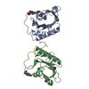
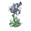
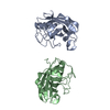
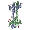
| Title | Crystal structure of BPSS1823, a Mip-like chaperone from Burkholderia pseudomallei | |||||||||
 Components Components | PEPTIDYL-PROLYL CIS-TRANS ISOMERASE Prolyl isomerase Prolyl isomerase | |||||||||
 Keywords Keywords | ISOMERASE / MIP / PPIASE / VIRULENCE | |||||||||
| Function / homology |  Function and homology informationpeptidylprolyl isomerase / peptidyl-prolyl cis-trans isomerase activity / protein folding Function and homology informationpeptidylprolyl isomerase / peptidyl-prolyl cis-trans isomerase activity / protein foldingSimilarity search - Function | |||||||||
| Biological species |  BURKHOLDERIA PSEUDOMALLEI (bacteria) BURKHOLDERIA PSEUDOMALLEI (bacteria) | |||||||||
| Method | X-RAY DIFFRACTION / SYNCHROTRON / MOLECULAR REPLACEMENT / Resolution: 0.91 Å | |||||||||
 Authors Authors | Norville, I.H. / O'Shea, K. / Sarkar-Tyson, M. / Harmer, N.J. | |||||||||
 Citation Citation | Journal: Biochem.J. / Year: 2011 Title: The Structure of a Burkholderia Pseudomallei Immunophilin-Inhibitor Complex Reveals New Approaches to Antimicrobial Development Authors: Norville, I.H. / O'Shea, K. / Sarkar-Tyson, M. / Zheng, S. / Titball, R.W. / Varani, G. / Harmer, N.J. | |||||||||
| History |
|
- Structure visualization
| Structure viewer | Molecule: MolmilJmol/JSmol |
|---|
- Downloads & links
-Download
| PDBx/mmCIF format | 2y78.cif.gz | 99.3 KB | Display | PDBx/mmCIF format |
|---|---|---|---|---|
| PDB format | pdb2y78.ent.gz | 76.7 KB | Display | PDB format |
| PDBx/mmJSON format | 2y78.json.gz | Tree view | PDBx/mmJSON format | |
| Others |  Other downloads Other downloads |
-Validation report
| Arichive directory | https://data.pdbj.org/pub/pdb/validation_reports/y7/2y78ftp://data.pdbj.org/pub/pdb/validation_reports/y7/2y78 | HTTPS FTP |
|---|
-Related structure data
| Related structure data |  2ke0C 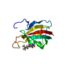 2ko7C  1fkbS  1rotS S: Starting model for refinement C: citing same article ( |
|---|---|
| Similar structure data |


-Links
 PDBj
PDBj

- Assembly
| Deposited unit | 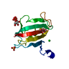
| ||||||||
|---|---|---|---|---|---|---|---|---|---|
| 1 | 
| ||||||||
| Unit cell |
| ||||||||
| Components on special symmetry positions |
|
-Components
| #1: Protein | Prolyl isomerase / PROLYL-PEPTIDE ISOMERASE Mass: 14112.774 Da / Num. of mol.: 1 Source method: isolated from a genetically manipulated source Source: (gene. exp.) BURKHOLDERIA PSEUDOMALLEI (bacteria) / Strain: K96243 / Production host: ESCHERICHIA COLI (E. coli) / Strain (production host): ROSETTA2 / References: UniProt: Q63J95, peptidylprolyl isomerase | ||||||
|---|---|---|---|---|---|---|---|
| #2: Chemical | Sulfate  Mass: 96.063 Da / Num. of mol.: 2 / Source method: obtained synthetically / Formula: SO4 Mass: 96.063 Da / Num. of mol.: 2 / Source method: obtained synthetically / Formula: SO4#3: Chemical | Glycerol  Mass: 92.094 Da / Num. of mol.: 2 / Source method: obtained synthetically / Formula: C3H8O3 Mass: 92.094 Da / Num. of mol.: 2 / Source method: obtained synthetically / Formula: C3H8O3#4: Chemical | ChemComp-CL / | Chloride  Mass: 35.453 Da / Num. of mol.: 1 / Source method: obtained synthetically / Formula: Cl Mass: 35.453 Da / Num. of mol.: 1 / Source method: obtained synthetically / Formula: Cl#5: Water | ChemComp-HOH / | Water Mass: 18.015 Da / Num. of mol.: 274 / Source method: isolated from a natural source / Formula: H2O Mass: 18.015 Da / Num. of mol.: 274 / Source method: isolated from a natural source / Formula: H2O |
-Experimental details
-Experiment
| Experiment | Method: X-RAY DIFFRACTION / Number of used crystals: 1 |
|---|
- Sample preparation
| Crystal | Density Matthews: 3.16 Å3/Da / Density % sol: 61.1 % / Description: NONE |
|---|---|
| Crystal grow | pH: 5.5 / Details: 1.1 M (NH4)2SO4, 0.05 M BIS-TRIS PH 5.5 |
-Data collection
| Diffraction | Mean temperature: 100 K |
|---|---|
| Diffraction source | Source: SYNCHROTRON / Site: Diamond  / Beamline: I02 / Wavelength: 0.861 / Beamline: I02 / Wavelength: 0.861 |
| Detector | Type: ADSC CCD / Detector: CCD / Date: Apr 23, 2009 / Details: MIRRORS |
| Radiation | Monochromator: SI CRYSTAL / Protocol: SINGLE WAVELENGTH / Monochromatic (M) / Laue (L): M / Scattering type: x-ray |
| Radiation wavelength | Wavelength: 0.861 Å / Relative weight: 1 |
| Reflection | Resolution: 0.91→40.3 Å / Num. obs: 125244 / % possible obs: 98.3 % / Observed criterion σ(I): 0 / Redundancy: 5.9 % / Biso Wilson estimate: 8.07 Å2 / Rmerge(I) obs: 0.08 / Net I/σ(I): 13.8 |
| Reflection shell | Resolution: 0.91→0.96 Å / Redundancy: 2.7 % / Rmerge(I) obs: 0.4 / Mean I/σ(I) obs: 2.2 / % possible all: 88.2 |
- Processing
| Software |
| |||||||||||||||||||||||||||||||||||||||||||||||||||||||||||||||||||||||||||||
|---|---|---|---|---|---|---|---|---|---|---|---|---|---|---|---|---|---|---|---|---|---|---|---|---|---|---|---|---|---|---|---|---|---|---|---|---|---|---|---|---|---|---|---|---|---|---|---|---|---|---|---|---|---|---|---|---|---|---|---|---|---|---|---|---|---|---|---|---|---|---|---|---|---|---|---|---|---|---|
| Refinement | Method to determine structure: MOLECULAR REPLACEMENT Starting model: PDB ENTRIES 1FKB AND 1ROT Resolution: 0.91→26.646 Å / SU ML: 0.08 / σ(F): 0.01 / Phase error: 8.42 / Stereochemistry target values: ML Details: RESIDUES BEFORE GLYCINE -8 ARE DISORDERED. G-8 TO H0 ARE ORDERED.
| |||||||||||||||||||||||||||||||||||||||||||||||||||||||||||||||||||||||||||||
| Solvent computation | Shrinkage radii: 0.9 Å / VDW probe radii: 1.11 Å / Solvent model: FLAT BULK SOLVENT MODEL / Bsol: 60 Å2 / ksol: 0.4 e/Å3 | |||||||||||||||||||||||||||||||||||||||||||||||||||||||||||||||||||||||||||||
| Displacement parameters | Biso mean: 14 Å2
| |||||||||||||||||||||||||||||||||||||||||||||||||||||||||||||||||||||||||||||
| Refinement step | Cycle: LAST / Resolution: 0.91→26.646 Å
| |||||||||||||||||||||||||||||||||||||||||||||||||||||||||||||||||||||||||||||
| Refine LS restraints |
| |||||||||||||||||||||||||||||||||||||||||||||||||||||||||||||||||||||||||||||
| LS refinement shell |
|
