Mass: 96.063 Da / Num. of mol.: 8 / Source method: obtained synthetically / Formula: SO4
Compound details
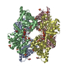
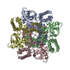
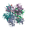
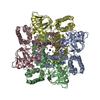
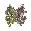
ENGINEERED RESIDUE IN CHAIN A, TYR 10 TO LYS ENGINEERED RESIDUE IN CHAIN B, TYR 10 TO LYS ...ENGINEERED RESIDUE IN CHAIN A, TYR 10 TO LYS ENGINEERED RESIDUE IN CHAIN B, TYR 10 TO LYS ENGINEERED RESIDUE IN CHAIN C, TYR 10 TO LYS ENGINEERED RESIDUE IN CHAIN D, TYR 10 TO LYS
-
Experimental details
-
Experiment
Experiment
Method: X-RAY DIFFRACTION / Number of used crystals: 1
-
Sample preparation
Crystal
Density Matthews: 2.44 Å3/Da / Density % sol: 48.9 % / Description: NONE
Crystal grow
Temperature: 293 K / Method: vapor diffusion, sitting drop / pH: 4.5 Details: AT 293 K UPON VAPOR DIFFUSION OF SITTING DROPS OF 1 MICRO-LITER PROTEIN SOLUTION (5 MG/ML PROTEIN, 25 MM TRIS-HCL PH 8.3, 50 MM NACL, 2.5 MM DTT, 0.25 MM UDP-GLCA, AND 0.5 MM OXIDIZED NAD), ...Details: AT 293 K UPON VAPOR DIFFUSION OF SITTING DROPS OF 1 MICRO-LITER PROTEIN SOLUTION (5 MG/ML PROTEIN, 25 MM TRIS-HCL PH 8.3, 50 MM NACL, 2.5 MM DTT, 0.25 MM UDP-GLCA, AND 0.5 MM OXIDIZED NAD), AND 1 MICRO-LITER WELL SOLUTION (200 MM AMMONIUM SULFATE, 100 MM SODIUM ACETATE PH 4.5, 11-14 % (W/V) PEG 4K, AND 50 MM NAF), EQUILIBRATED AGAINST 500 MICRO-LITER PRECIPITATION SOLUTION IN THE WELL.
Protocol: SINGLE WAVELENGTH / Monochromatic (M) / Laue (L): M / Scattering type: x-ray
Radiation wavelength
Wavelength: 0.9334 Å / Relative weight: 1
Reflection
Resolution: 2.8→57.83 Å / Num. obs: 50832 / % possible obs: 99.8 % / Observed criterion σ(I): 0 / Redundancy: 4.1 % / Biso Wilson estimate: 27.74 Å2 / Rmerge(I) obs: 0.22 / Net I/σ(I): 2.8
Reflection shell
Resolution: 2.8→2.95 Å / Redundancy: 4.1 % / Rmerge(I) obs: 0.44 / Mean I/σ(I) obs: 1.7 / % possible all: 100
-
Processing
Software
Name
Version
Classification
PHENIX
(PHENIX.REFINE)
refinement
MOSFLM
datareduction
SCALA
datascaling
PHASER
phasing
Refinement
Method to determine structure: MOLECULAR REPLACEMENT Starting model: NATIVE BCEC WITH TYR 10 TRUNCATED TO ALA Resolution: 2.8→54.63 Å / SU ML: 1.02 / σ(F): 1.36 / Phase error: 16.16 / Stereochemistry target values: ML
Rfactor
Num. reflection
% reflection
Rfree
0.264
2576
5.1 %
Rwork
0.212
-
-
obs
0.21
50791
99.6 %
Solvent computation
Shrinkage radii: 0.9 Å / VDW probe radii: 1.11 Å / Solvent model: FLAT BULK SOLVENT MODEL / Bsol: 13.37 Å2 / ksol: 0.36 e/Å3
Displacement parameters
Biso mean: 28.8 Å2
Baniso -1
Baniso -2
Baniso -3
1-
1.8056 Å2
0 Å2
0 Å2
2-
-
-7.5775 Å2
0 Å2
3-
-
-
0.9821 Å2
Refinement step
Cycle: LAST / Resolution: 2.8→54.63 Å
Protein
Nucleic acid
Ligand
Solvent
Total
Num. atoms
13937
0
188
0
14125
Refine LS restraints
Refine-ID
Type
Dev ideal
Number
X-RAY DIFFRACTION
f_bond_d
0.013
14387
X-RAY DIFFRACTION
f_angle_d
1.416
19503
X-RAY DIFFRACTION
f_dihedral_angle_d
17.901
5287
X-RAY DIFFRACTION
f_chiral_restr
0.098
2166
X-RAY DIFFRACTION
f_plane_restr
0.006
2558
Refine LS restraints NCS
Ens-ID
Dom-ID
Auth asym-ID
Number
Refine-ID
Type
Rms dev position (Å)
1
1
A
3455
X-RAY DIFFRACTION
POSITIONAL
1
2
B
3455
X-RAY DIFFRACTION
POSITIONAL
0.083
1
3
C
3455
X-RAY DIFFRACTION
POSITIONAL
0.099
1
4
D
3455
X-RAY DIFFRACTION
POSITIONAL
0.091
LS refinement shell
Resolution (Å)
Rfactor Rfree
Num. reflection Rfree
Rfactor Rwork
Num. reflection Rwork
Refine-ID
% reflection obs (%)
2.8-2.8539
0.328
132
0.2494
2625
X-RAY DIFFRACTION
100
2.8539-2.9121
0.3033
135
0.2439
2690
X-RAY DIFFRACTION
100
2.9121-2.9754
0.3003
156
0.2499
2626
X-RAY DIFFRACTION
100
2.9754-3.0446
0.304
152
0.2456
2644
X-RAY DIFFRACTION
100
3.0446-3.1208
0.3094
142
0.2589
2667
X-RAY DIFFRACTION
100
3.1208-3.2051
0.3115
146
0.2511
2643
X-RAY DIFFRACTION
100
3.2051-3.2994
0.3211
143
0.2292
2656
X-RAY DIFFRACTION
100
3.2994-3.4059
0.2691
152
0.2322
2657
X-RAY DIFFRACTION
100
3.4059-3.5276
0.299
135
0.2078
2671
X-RAY DIFFRACTION
100
3.5276-3.6689
0.2515
150
0.2025
2648
X-RAY DIFFRACTION
100
3.6689-3.8358
0.2482
152
0.188
2661
X-RAY DIFFRACTION
100
3.8358-4.038
0.2282
128
0.1781
2729
X-RAY DIFFRACTION
100
4.038-4.2909
0.1873
139
0.1708
2665
X-RAY DIFFRACTION
100
4.2909-4.622
0.2146
138
0.1593
2691
X-RAY DIFFRACTION
99
4.622-5.0868
0.2055
142
0.1539
2699
X-RAY DIFFRACTION
99
5.0868-5.8222
0.2284
140
0.1734
2707
X-RAY DIFFRACTION
99
5.8222-7.3325
0.2463
134
0.1886
2752
X-RAY DIFFRACTION
99
7.3325-54.6353
0.2066
160
0.194
2783
X-RAY DIFFRACTION
97
Refinement TLS params.
Method: refined / Refine-ID: X-RAY DIFFRACTION
ID
L11 (°2)
L12 (°2)
L13 (°2)
L22 (°2)
L23 (°2)
L33 (°2)
S11 (Å °)
S12 (Å °)
S13 (Å °)
S21 (Å °)
S22 (Å °)
S23 (Å °)
S31 (Å °)
S32 (Å °)
S33 (Å °)
T11 (Å2)
T12 (Å2)
T13 (Å2)
T22 (Å2)
T23 (Å2)
T33 (Å2)
Origin x (Å)
Origin y (Å)
Origin z (Å)
1
0.1939
-0.1204
-0.1099
0.0916
0.059
0.1016
-0.0315
0.013
-0.0978
0.0427
-0.0181
0.0306
-0.0291
-0.0264
-0.0391
0.034
0.0428
0.0152
-0.036
-0.0006
0.0248
-22.4269
-14.2568
48.0535
2
0.1194
-0.0068
-0.0067
0.0864
-0.0479
0.0767
0.0405
-0.0138
0.0865
-0.0802
0.0242
0.0703
0.0594
0.0546
0.0141
0.0915
0.0221
-0.025
0.0669
0.0006
0.0596
-23.9407
-26.8491
16.688
3
0.0775
-0.0999
0.0879
0.1015
-0.1159
0.1233
0.0307
0.2161
0.1151
-0.0756
-0.254
-0.1182
0.099
0.2158
-0.2191
-0.0094
0.163
0.0255
0.2403
0.2596
0.0078
-37.8466
4.3915
-7.7085
4
0.1994
-0.0163
0.0185
0.0496
-0.0105
-0.0029
0.0231
0.0188
0.0995
-0.0618
-0.0366
0.0351
0.0093
-0.0085
0.0447
-0.1711
0.0407
-0.0013
0.0345
-0.004
0.0844
-50.8694
-0.059
22.1295
5
0.012
0.0619
-0.0635
0.1499
-0.1271
0.1432
-0.0485
-0.0187
-0.0205
-0.1023
-0.0894
-0.1117
0.0589
0.0252
-0.188
0.0234
-0.0231
0.1364
-0.0374
0.1109
-0.0492
3.7906
-4.2108
7.559
6
0.0456
0.0217
-0.0374
0.0671
0.0125
0.1684
-0.0098
-0.0211
0.0445
-0.0496
-0.0078
0.028
-0.0097
0.0877
-0.0296
0.0122
-0.0079
-0.0174
0.0501
0.0078
0.0061
3.6484
10.027
37.8405
7
0.0502
0.0134
0.0297
0.0378
0.0724
0.1133
-0.1648
-0.1307
-0.0436
0.1068
0.0339
0.2174
0.0443
-0.0113
-0.0308
-0.004
0.1762
0.1494
-0.0222
-0.007
0.1876
-33.7425
28.0911
42.3305
8
0.0009
-0.0211
-0.0038
0.0723
-0.0115
-0.0118
-0.0027
-0.0283
-0.0308
-0.0728
0.0063
0.0501
-0.0184
0.0067
-0.0172
0.0504
0.0224
-0.0598
-0.0675
0.0459
-0.0178
-17.3854
31.3719
13.0497
Refinement TLS group
ID
Refine-ID
Refine TLS-ID
Selection details
1
X-RAY DIFFRACTION
1
(CHAINAANDRESID0:203)
2
X-RAY DIFFRACTION
2
(CHAINAANDRESID204:456)
3
X-RAY DIFFRACTION
3
(CHAINBANDRESID0:195)
4
X-RAY DIFFRACTION
4
(CHAINBANDRESID196:453)
5
X-RAY DIFFRACTION
5
(CHAINCANDRESID0:201)
6
X-RAY DIFFRACTION
6
(CHAINCANDRESID202:453)
7
X-RAY DIFFRACTION
7
(CHAINDANDRESID0:201)
8
X-RAY DIFFRACTION
8
(CHAINDANDRESID202:455)
+
About Yorodumi
-
News
-
Feb 9, 2022. New format data for meta-information of EMDB entries
New format data for meta-information of EMDB entries
Version 3 of the EMDB header file is now the official format.
The previous official version 1.9 will be removed from the archive.
In the structure databanks used in Yorodumi, some data are registered as the other names, "COVID-19 virus" and "2019-nCoV". Here are the details of the virus and the list of structure data.
Jan 31, 2019. EMDB accession codes are about to change! (news from PDBe EMDB page)
EMDB accession codes are about to change! (news from PDBe EMDB page)
The allocation of 4 digits for EMDB accession codes will soon come to an end. Whilst these codes will remain in use, new EMDB accession codes will include an additional digit and will expand incrementally as the available range of codes is exhausted. The current 4-digit format prefixed with “EMD-” (i.e. EMD-XXXX) will advance to a 5-digit format (i.e. EMD-XXXXX), and so on. It is currently estimated that the 4-digit codes will be depleted around Spring 2019, at which point the 5-digit format will come into force.
The EM Navigator/Yorodumi systems omit the EMD- prefix.
Related info.:Q: What is EMD? / ID/Accession-code notation in Yorodumi/EM Navigator
Yorodumi is a browser for structure data from EMDB, PDB, SASBDB, etc.
This page is also the successor to EM Navigator detail page, and also detail information page/front-end page for Omokage search.
The word "yorodu" (or yorozu) is an old Japanese word meaning "ten thousand". "mi" (miru) is to see.
Related info.:EMDB / PDB / SASBDB / Comparison of 3 databanks / Yorodumi Search / Aug 31, 2016. New EM Navigator & Yorodumi / Yorodumi Papers / Jmol/JSmol / Function and homology information / Changes in new EM Navigator and Yorodumi
 Movie
Movie Controller


 Open data
 Basic information
 Components
 Keywords
 OXIDOREDUCTASE /
  Function and homology information
 Authors
 Citation
 Structure visualization
 Downloads & links
 Other downloads
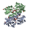


 PDBj
PDBj


 Assembly
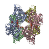
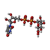
 Mass: 580.285 Da / Num. of mol.: 4 / Source method: obtained synthetically / Formula: C15H22N2O18P2
 Mass: 96.063 Da / Num. of mol.: 8 / Source method: obtained synthetically / Formula: SO4
 Sample preparation
 / Beamline: ID14-1 / Wavelength: 0.9334
  Processing