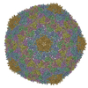
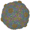
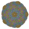
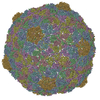
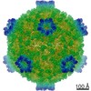
Journal: J Biol Chem / Year: 2011 Title: Molecular rearrangements involved in the capsid shell maturation of bacteriophage T7. Authors: Alina Ionel / Javier A Velázquez-Muriel / Daniel Luque / Ana Cuervo / José R Castón / José M Valpuesta / Jaime Martín-Benito / José L Carrascosa / Abstract: Maturation of dsDNA bacteriophages involves assembling the virus prohead from a limited set of structural components followed by rearrangements required for the stability that is necessary for ...Maturation of dsDNA bacteriophages involves assembling the virus prohead from a limited set of structural components followed by rearrangements required for the stability that is necessary for infecting a host under challenging environmental conditions. Here, we determine the mature capsid structure of T7 at 1 nm resolution by cryo-electron microscopy and compare it with the prohead to reveal the molecular basis of T7 shell maturation. The mature capsid presents an expanded and thinner shell, with a drastic rearrangement of the major protein monomers that increases in their interacting surfaces, in turn resulting in a new bonding lattice. The rearrangements include tilting, in-plane rotation, and radial expansion of the subunits, as well as a relative bending of the A- and P-domains of each subunit. The unique features of this shell transformation, which does not employ the accessory proteins, inserted domains, or molecular interactions observed in other phages, suggest a simple capsid assembling strategy that may have appeared early in the evolution of these viruses.
A: MAJOR CAPSID PROTEIN 10A B: MAJOR CAPSID PROTEIN 10A C: MAJOR CAPSID PROTEIN 10A D: MAJOR CAPSID PROTEIN 10A E: MAJOR CAPSID PROTEIN 10A F: MAJOR CAPSID PROTEIN 10A G: MAJOR CAPSID PROTEIN 10A
A: MAJOR CAPSID PROTEIN 10A B: MAJOR CAPSID PROTEIN 10A C: MAJOR CAPSID PROTEIN 10A D: MAJOR CAPSID PROTEIN 10A E: MAJOR CAPSID PROTEIN 10A F: MAJOR CAPSID PROTEIN 10A G: MAJOR CAPSID PROTEIN 10A
Idetical with deposited unit in distinct coordinate
icosahedral asymmetric unit
Type
Name
Symmetry operation
Number
point symmetry operation
1
3
A: MAJOR CAPSID PROTEIN 10A B: MAJOR CAPSID PROTEIN 10A C: MAJOR CAPSID PROTEIN 10A D: MAJOR CAPSID PROTEIN 10A E: MAJOR CAPSID PROTEIN 10A F: MAJOR CAPSID PROTEIN 10A G: MAJOR CAPSID PROTEIN 10A
x 5
icosahedral pentamer
1.28 MDa, 35 polymers
Theoretical mass
Number of molelcules
Total (without water)
1,280,637
35
Polymers
1,280,637
35
Non-polymers
0
0
Water
0
Type
Name
Symmetry operation
Number
point symmetry operation
5
4
A: MAJOR CAPSID PROTEIN 10A B: MAJOR CAPSID PROTEIN 10A C: MAJOR CAPSID PROTEIN 10A D: MAJOR CAPSID PROTEIN 10A E: MAJOR CAPSID PROTEIN 10A F: MAJOR CAPSID PROTEIN 10A G: MAJOR CAPSID PROTEIN 10A
x 6
icosahedral 23 hexamer
1.54 MDa, 42 polymers
Theoretical mass
Number of molelcules
Total (without water)
1,536,764
42
Polymers
1,536,764
42
Non-polymers
0
0
Water
0
Type
Name
Symmetry operation
Number
point symmetry operation
6
5
Idetical with deposited unit in distinct coordinate
icosahedral asymmetric unit, std point frame
Type
Name
Symmetry operation
Number
transform to point frame
1
Symmetry
Point symmetry: (Schoenflies symbol: I (icosahedral))
-
Components
#1: Protein
MAJORCAPSIDPROTEIN10A / BACTERIOPHAGE T7
Mass: 36589.625 Da / Num. of mol.: 7 / Source method: isolated from a natural source / Source: (natural) ENTEROBACTERIA PHAGE T7 (virus) / References: UniProt: P19726
-
Experimental details
-
Experiment
Experiment
Method: ELECTRON MICROSCOPY
EM experiment
Aggregation state: PARTICLE / 3D reconstruction method: single particle reconstruction
-
Sample preparation
Component
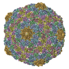
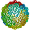
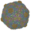
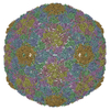
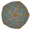
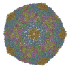
Name: PHAGE T7 EMPTY HEAD / Type: VIRUS
Buffer solution
Name: 50 MM TRIS-HCL, PH 7.8, 10 MM MGCL2, 0.1 M NACL / pH: 7.8 / Details: 50 MM TRIS-HCL, PH 7.8, 10 MM MGCL2, 0.1 M NACL
Specimen
Embedding applied: NO / Shadowing applied: NO / Staining applied: NO / Vitrification applied: YES
Electron source: FIELD EMISSION GUN / Accelerating voltage: 200 kV / Illumination mode: FLOOD BEAM
Electron lens
Mode: BRIGHT FIELDBright-field microscopy / Nominal magnification: 50000 X
Image recording
Film or detector model: KODAK SO-163 FILM
-
Processing
EM software
Name: Xmipp / Category: 3D reconstruction
Symmetry
Point symmetry: I (icosahedral)
3D reconstruction
Resolution: 10.8 Å / Num. of particles: 5100 / Actual pixel size: 1.4 Å Details: SUBMISSION BASED ON EXPERIMENTAL DATA FROM EMDB EMD-1810. (DEPOSITION ID: 7638). Symmetry type: POINT
In the structure databanks used in Yorodumi, some data are registered as the other names, "COVID-19 virus" and "2019-nCoV". Here are the details of the virus and the list of structure data.
Jan 31, 2019. EMDB accession codes are about to change! (news from PDBe EMDB page)
EMDB accession codes are about to change! (news from PDBe EMDB page)
The allocation of 4 digits for EMDB accession codes will soon come to an end. Whilst these codes will remain in use, new EMDB accession codes will include an additional digit and will expand incrementally as the available range of codes is exhausted. The current 4-digit format prefixed with “EMD-” (i.e. EMD-XXXX) will advance to a 5-digit format (i.e. EMD-XXXXX), and so on. It is currently estimated that the 4-digit codes will be depleted around Spring 2019, at which point the 5-digit format will come into force.
The EM Navigator/Yorodumi systems omit the EMD- prefix.
Related info.:Q: What is EMD? / ID/Accession-code notation in Yorodumi/EM Navigator
Yorodumi is a browser for structure data from EMDB, PDB, SASBDB, etc.
This page is also the successor to EM Navigator detail page, and also detail information page/front-end page for Omokage search.
The word "yorodu" (or yorozu) is an old Japanese word meaning "ten thousand". "mi" (miru) is to see.
Related info.:EMDB / PDB / SASBDB / Comparison of 3 databanks / Yorodumi Search / Aug 31, 2016. New EM Navigator & Yorodumi / Yorodumi Papers / Jmol/JSmol / Function and homology information / Changes in new EM Navigator and Yorodumi
 Movie
Movie Controller


 Open data
 Basic information
 Components
 Keywords
 VIRUS / CAPSID MATURATION / MORPHOGENETIC INTERMEDIATE
 Function and homology information

 Authors
 Citation
 Structure visualization
 Downloads & links
 Other downloads
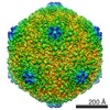
 PDBj
PDBj Assembly


 Sample preparation
 Electron microscopy imaging
 Processing