[English] 日本語
Yorodumi
- PDB-2xvl: crystal structure of alpha-xylosidase (GH31) from Cellvibrio japo... -

+
Open data


ID or keywords:

Loading...

-
Basic information

Entry
Database: PDB / ID: 2xvl
Titlecrystal structure of alpha-xylosidase (GH31) from Cellvibrio japonicus in complex with Pentaerythritol propoxylate (5 4 PO OH)
ComponentsALPHA-XYLOSIDASE, PUTATIVE, XYL31A
Keywords
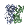
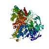
HYDROLASE / GLYCOSYL HYDROLASE FAMILY 31 / (BETA/ALPHA)8 BARREL
Function / homology
Function and homology information


Hydrolases; Glycosylases; Glycosidases, i.e. enzymes that hydrolyse O- and S-glycosyl compounds / hydrolase activity, hydrolyzing O-glycosyl compounds / carbohydrate binding / carbohydrate metabolic process
Similarity search - Function
Jelly Rolls - #380 / : / Domain of unknown function DUF5110 / Domain of unknown function (DUF5110) / glycosyl hydrolase (family 31) / PA14/GLEYA domain / PA14 domain profile. / Glycoside hydrolase family 31, N-terminal domain / Glycosyl hydrolase 31 N-terminal galactose mutarotase-like domain / PA14 ...Jelly Rolls - #380 / : / Domain of unknown function DUF5110 / Domain of unknown function (DUF5110) / glycosyl hydrolase (family 31) / PA14/GLEYA domain / PA14 domain profile. / Glycoside hydrolase family 31, N-terminal domain / Glycosyl hydrolase 31 N-terminal galactose mutarotase-like domain / PA14 / PA14 domain / : / Glycosyl hydrolase family 31 C-terminal domain / Glycoside hydrolase family 31 / Glycosyl hydrolases family 31 TIM-barrel domain / Galactose mutarotase-like domain superfamily / Golgi alpha-mannosidase II / Glycosyl hydrolase, all-beta / Glycosidases / Glycoside hydrolase superfamily / Jelly Rolls / TIM Barrel / Alpha-Beta Barrel / Immunoglobulin-like / Sandwich / Mainly Beta / Alpha Beta
Similarity search - Domain/homology
NICKEL (II) ION / Chem-PXN / Alpha-xylosidase, putative, xyl31A
Similarity search - Component
Biological speciesCELLVIBRIO JAPONICUS (bacteria)
MethodX-RAY DIFFRACTION / SYNCHROTRON / MOLECULAR REPLACEMENT / Resolution: 2.3 Å
AuthorsLarsbrink, J. / Izumi, A. / Ibatullin, F. / Nakhai, A. / Gilbert, H.J. / Davies, G.J. / Brumer, H.
CitationJournal: Biochem.J. / Year: 2011
Title: Structural and Enzymatic Characterisation of a Glycoside Hydrolase Family 31 Alpha-Xylosidase from Cellvibrio Japonicus Involved in Xyloglucan Saccharification.
Authors: Larsbrink, J. / Izumi, A. / Ibatullin, F. / Nakhai, A. / Gilbert, H.J. / Davies, G.J. / Brumer, H.
History
DepositionOct 26, 2010Deposition site: PDBE / Processing site: PDBE
Revision 1.0Apr 13, 2011Provider: repository / Type: Initial release
Revision 1.1Jun 2, 2011Group: Version format compliance
Revision 1.2Jul 13, 2011Group: Version format compliance
Revision 1.3Dec 20, 2023Group: Data collection / Database references ...Data collection / Database references / Derived calculations / Other / Refinement description
Category: chem_comp_atom / chem_comp_bond ...chem_comp_atom / chem_comp_bond / database_2 / pdbx_database_status / pdbx_initial_refinement_model / pdbx_struct_conn_angle / struct_conn / struct_site
Item: _database_2.pdbx_DOI / _database_2.pdbx_database_accession ..._database_2.pdbx_DOI / _database_2.pdbx_database_accession / _pdbx_database_status.status_code_sf / _pdbx_struct_conn_angle.ptnr1_auth_comp_id / _pdbx_struct_conn_angle.ptnr1_auth_seq_id / _pdbx_struct_conn_angle.ptnr1_label_asym_id / _pdbx_struct_conn_angle.ptnr1_label_atom_id / _pdbx_struct_conn_angle.ptnr1_label_comp_id / _pdbx_struct_conn_angle.ptnr1_label_seq_id / _pdbx_struct_conn_angle.ptnr2_auth_seq_id / _pdbx_struct_conn_angle.ptnr2_label_asym_id / _pdbx_struct_conn_angle.ptnr3_auth_comp_id / _pdbx_struct_conn_angle.ptnr3_auth_seq_id / _pdbx_struct_conn_angle.ptnr3_label_asym_id / _pdbx_struct_conn_angle.ptnr3_label_atom_id / _pdbx_struct_conn_angle.ptnr3_label_comp_id / _pdbx_struct_conn_angle.ptnr3_label_seq_id / _pdbx_struct_conn_angle.value / _struct_conn.pdbx_dist_value / _struct_conn.ptnr1_auth_comp_id / _struct_conn.ptnr1_auth_seq_id / _struct_conn.ptnr1_label_asym_id / _struct_conn.ptnr1_label_atom_id / _struct_conn.ptnr1_label_comp_id / _struct_conn.ptnr1_label_seq_id / _struct_conn.ptnr2_auth_comp_id / _struct_conn.ptnr2_auth_seq_id / _struct_conn.ptnr2_label_asym_id / _struct_conn.ptnr2_label_atom_id / _struct_conn.ptnr2_label_comp_id / _struct_conn.ptnr2_label_seq_id / _struct_site.pdbx_auth_asym_id / _struct_site.pdbx_auth_comp_id / _struct_site.pdbx_auth_seq_id

-
Structure visualization

Structure viewerMolecule:
MolmilJmol/JSmol

Downloads & links

-
Assembly

Deposited unit
A: ALPHA-XYLOSIDASE, PUTATIVE, XYL31A
hetero molecules


Theoretical massNumber of molelcules
Total (without water)116,28515
Polymers115,2041
Non-polymers1,08114
Water12,863714
1


  • Idetical with deposited unit
  • defined by author&software
TypeNameSymmetry operationNumber
identity operation1_555x,y,z1
MethodPISA
Unit cell
Length a, b, c (Å)157.653, 157.653, 226.688
Angle α, β, γ (deg.)90.00, 90.00, 120.00
Int Tables number182
Space group name H-MP6322
Components on special symmetry positions
IDModelComponents
11A-1995-

CL

21A-2400-

HOH

-
Components

-
Protein , 1 types, 1 molecules A

#1: Protein ALPHA-XYLOSIDASE, PUTATIVE, XYL31A / ALPHA XYLOSIDASE


Mass: 115203.914 Da / Num. of mol.: 1
Source method: isolated from a genetically manipulated source
Source: (gene. exp.) CELLVIBRIO JAPONICUS (bacteria) / Production host: ESCHERICHIA COLI (E. coli) / Strain (production host): BL21(DE3)
References: UniProt: B3PBD9, Hydrolases; Glycosylases; Glycosidases, i.e. enzymes that hydrolyse O- and S-glycosyl compounds

-
Non-polymers , 5 types, 728 molecules

#2: Chemical ChemComp-NI / NICKEL (II) ION / Nickel


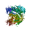
Mass: 58.693 Da / Num. of mol.: 3 / Source method: obtained synthetically / Formula: Ni
#3: Chemical ChemComp-SO4 / SULFATE ION / Sulfate


Mass: 96.063 Da / Num. of mol.: 3 / Source method: obtained synthetically / Formula: SO4
#4: Chemical
ChemComp-CL / CHLORIDE ION / Chloride


Mass: 35.453 Da / Num. of mol.: 7 / Source method: obtained synthetically / Formula: Cl
#5: Chemical ChemComp-PXN / (2S)-1-[3-{[(2R)-2-hydroxypropyl]oxy}-2,2-bis({[(2R)-2-hydroxypropyl]oxy}methyl)propoxy]propan-2-ol / PENTAERYTHRITOL PROPOXYLATE (5/4 PO/OH)


Mass: 368.463 Da / Num. of mol.: 1 / Source method: obtained synthetically / Formula: C17H36O8
#6: Water ChemComp-HOH / water / Water


Mass: 18.015 Da / Num. of mol.: 714 / Source method: isolated from a natural source / Formula: H2O

-
Details

Nonpolymer detailsPENTAERYTHRITOL PROPOXYLATE (5 4 PO OH) (54P): AVERAGE MOLECULAR WEIGHT 426

-
Experimental details

-
Experiment

ExperimentMethod: X-RAY DIFFRACTION / Number of used crystals: 1

-
Sample preparation

CrystalDensity Matthews: 3.53 Å3/Da / Density % sol: 65.15 %
Crystal growpH: 7
Details: 40 % PENTAERYTHRITOL PROPOXYLATE (5/4 PO/OH), 0.1 M BIS-TRIS (PH 7.0), 5 MM NI SO4

-
Data collection

DiffractionMean temperature: 100 K
Diffraction sourceSource: SYNCHROTRON / Site: ESRF / Beamline: ID29 / Wavelength: 0.9334
DetectorType: ADSC QUANTUM 315r / Detector: CCD / Date: Sep 26, 2009 / Details: MIRRORS
RadiationMonochromator: DIAMOND (111),GE(220) / Protocol: SINGLE WAVELENGTH / Monochromatic (M) / Laue (L): M / Scattering type: x-ray
Radiation wavelengthWavelength: 0.9334 Å / Relative weight: 1
ReflectionResolution: 2.3→58.5 Å / Num. obs: 74143 / % possible obs: 100 % / Observed criterion σ(I): -3 / Redundancy: 11.3 % / Biso Wilson estimate: 24.33 Å2 / Rmerge(I) obs: 0.17 / Net I/σ(I): 16.3
Reflection shellResolution: 2.3→2.42 Å / Redundancy: 4.5 % / Rmerge(I) obs: 0.47 / Mean I/σ(I) obs: 7.5 / % possible all: 100

-
Processing

Software
NameVersionClassification
PHENIX(PHENIX.REFINE)refinement
MOSFLMdata reduction
SCALAdata scaling
BALBESphasing
RefinementMethod to determine structure: MOLECULAR REPLACEMENT
Starting model: PDB ENTRY 2G3M
Resolution: 2.3→54.548 Å / SU ML: 0.22 / σ(F): 1.38 / Phase error: 16.07 / Stereochemistry target values: ML
Details: DISORDERED SIDE CHAINS WERE MODELED WITH ZERO-OCCUPANCY.
RfactorNum. reflection% reflection
Rfree0.1853 3736 5 %
Rwork0.145 --
obs0.147 74143 99.99 %
Solvent computationShrinkage radii: 0.9 Å / VDW probe radii: 1.11 Å / Solvent model: FLAT BULK SOLVENT MODEL / Bsol: 34.4 Å2 / ksol: 0.322 e/Å3
Displacement parameters
Baniso -1Baniso -2Baniso -3
1-2.8439 Å20 Å20 Å2
2--2.8439 Å20 Å2
3----5.6878 Å2
Refinement stepCycle: LAST / Resolution: 2.3→54.548 Å
ProteinNucleic acidLigandSolventTotal
Num. atoms7572 0 50 714 8336
Refine LS restraints
Refine-IDTypeDev idealNumber
X-RAY DIFFRACTIONf_bond_d0.0077830
X-RAY DIFFRACTIONf_angle_d1.04310638
X-RAY DIFFRACTIONf_dihedral_angle_d17.1832793
X-RAY DIFFRACTIONf_chiral_restr0.071101
X-RAY DIFFRACTIONf_plane_restr0.0041391
LS refinement shell
Resolution (Å)Rfactor RfreeNum. reflection RfreeRfactor RworkNum. reflection RworkRefine-ID% reflection obs (%)
2.3-2.38220.25893370.18936956X-RAY DIFFRACTION100
2.3822-2.47760.22563820.16756883X-RAY DIFFRACTION100
2.4776-2.59040.20783510.16166968X-RAY DIFFRACTION100
2.5904-2.72690.193600.15236974X-RAY DIFFRACTION100
2.7269-2.89780.22123850.15196942X-RAY DIFFRACTION100
2.8978-3.12150.18673870.15316961X-RAY DIFFRACTION100
3.1215-3.43560.19233880.14457011X-RAY DIFFRACTION100
3.4356-3.93260.17174010.13727044X-RAY DIFFRACTION100
3.9326-4.95410.13193650.10797159X-RAY DIFFRACTION100
4.9541-54.56360.17563800.1457509X-RAY DIFFRACTION100
Refinement TLS params.

Method: refined / Refine-ID: X-RAY DIFFRACTION

IDL112)L122)L132)L222)L232)L332)S11 (Å °)S12 (Å °)S13 (Å °)S21 (Å °)S22 (Å °)S23 (Å °)S31 (Å °)S32 (Å °)S33 (Å °)T112)T122)T132)T222)T232)T332)Origin x (Å)Origin y (Å)Origin z (Å)
10.2192-0.2783-0.05150.45010.24530.63680.01960.04370.01750.02880.1013-0.21140.20070.4304-0.12420.05530.134-0.00450.4744-0.09250.175662.829573.097621.6658
20.2451-0.064-0.07010.4870.15790.53920.0690.02040.1376-0.01910.0449-0.2023-0.13450.2907-0.10890.0948-0.0890.04590.3061-0.08520.241552.9523106.617833.5811
30.3183-0.14170.02270.38220.04610.40440.0038-0.09330.03640.0960.0672-0.0881-0.01440.1175-0.06490.0470.0365-0.00260.1548-0.0370.03932.255387.390241.9725
40.4397-0.2381-0.03490.3988-0.00760.58680.03340.03250.0907-0.01310.0185-0.1003-0.04840.1332-0.05160.03260.00740.02930.1087-0.01460.058128.892192.446323.7425
50.4442-0.1952-0.10080.3042-0.06110.07460.05790.2535-0.0799-0.1881-0.00050.07370.0466-0.1553-0.0270.12730.0576-0.02840.2108-0.03330.017313.689884.53752.775
Refinement TLS group
IDRefine-IDRefine TLS-IDSelection details
1X-RAY DIFFRACTION1(CHAIN A AND RESID 45:142)
2X-RAY DIFFRACTION2(CHAIN A AND RESID 143:384)
3X-RAY DIFFRACTION3(CHAIN A AND RESID 385:578)
4X-RAY DIFFRACTION4(CHAIN A AND RESID 579:950)
5X-RAY DIFFRACTION5(CHAIN A AND RESID 951:988)

+
About Yorodumi

-
News

-
Feb 9, 2022. New format data for meta-information of EMDB entries

New format data for meta-information of EMDB entries

  • Version 3 of the EMDB header file is now the official format.
  • The previous official version 1.9 will be removed from the archive.

Related info.:EMDB header

External links:wwPDB to switch to version 3 of the EMDB data model

-
Aug 12, 2020. Covid-19 info

Covid-19 info

URL: https://pdbj.org/emnavi/covid19.php

New page: Covid-19 featured information page in EM Navigator.

Related info.:Covid-19 info / Mar 5, 2020. Novel coronavirus structure data

+
Mar 5, 2020. Novel coronavirus structure data

Novel coronavirus structure data

Related info.:Yorodumi Speices / Aug 12, 2020. Covid-19 info

External links:COVID-19 featured content - PDBj / Molecule of the Month (242):Coronavirus Proteases

+
Jan 31, 2019. EMDB accession codes are about to change! (news from PDBe EMDB page)

EMDB accession codes are about to change! (news from PDBe EMDB page)

  • The allocation of 4 digits for EMDB accession codes will soon come to an end. Whilst these codes will remain in use, new EMDB accession codes will include an additional digit and will expand incrementally as the available range of codes is exhausted. The current 4-digit format prefixed with “EMD-” (i.e. EMD-XXXX) will advance to a 5-digit format (i.e. EMD-XXXXX), and so on. It is currently estimated that the 4-digit codes will be depleted around Spring 2019, at which point the 5-digit format will come into force.
  • The EM Navigator/Yorodumi systems omit the EMD- prefix.

Related info.:Q: What is EMD? / ID/Accession-code notation in Yorodumi/EM Navigator

External links:EMDB Accession Codes are Changing Soon! / Contact to PDBj

+
Jul 12, 2017. Major update of PDB

Major update of PDB

  • wwPDB released updated PDB data conforming to the new PDBx/mmCIF dictionary.
  • This is a major update changing the version number from 4 to 5, and with Remediation, in which all the entries are updated.
  • In this update, many items about electron microscopy experimental information are reorganized (e.g. em_software).
  • Now, EM Navigator and Yorodumi are based on the updated data.

External links:wwPDB Remediation / Enriched Model Files Conforming to OneDep Data Standards Now Available in the PDB FTP Archive

-
Yorodumi

Thousand views of thousand structures

  • Yorodumi is a browser for structure data from EMDB, PDB, SASBDB, etc.
  • This page is also the successor to EM Navigator detail page, and also detail information page/front-end page for Omokage search.
  • The word "yorodu" (or yorozu) is an old Japanese word meaning "ten thousand". "mi" (miru) is to see.

Related info.:EMDB / PDB / SASBDB / Comparison of 3 databanks / Yorodumi Search / Aug 31, 2016. New EM Navigator & Yorodumi / Yorodumi Papers / Jmol/JSmol / Function and homology information / Changes in new EM Navigator and Yorodumi

Read more