 Movie
Movie Controller

[English] 日本語
 Yorodumi
- PDB-2xsz: The dodecameric human RuvBL1:RuvBL2 complex with truncated domains II -
+ Open data

- Basic information
| Entry | Database: PDB / ID: 2xsz | ||||||
|---|---|---|---|---|---|---|---|
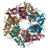
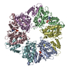
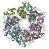
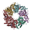
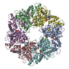
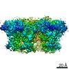
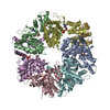
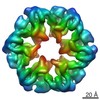
| Title | The dodecameric human RuvBL1:RuvBL2 complex with truncated domains II | ||||||
 Components Components |
| ||||||
 Keywords Keywords |  HYDROLASE / AAA+ PROTEINS / HELICASE / CHROMATIN REMODELLING HYDROLASE / AAA+ PROTEINS / HELICASE / CHROMATIN REMODELLING | ||||||
| Function / homology |  Function and homology information Function and homology informationpromoter-enhancer loop anchoring activity / regulation of DNA strand elongation / positive regulation of telomere maintenance in response to DNA damage / establishment of protein localization to chromatin / R2TP complex / Swr1 complex / dynein axonemal particle / RPAP3/R2TP/prefoldin-like complex / regulation of double-strand break repair / positive regulation of telomerase RNA localization to Cajal body ...promoter-enhancer loop anchoring activity / regulation of DNA strand elongation / positive regulation of telomere maintenance in response to DNA damage / establishment of protein localization to chromatin / R2TP complex / Swr1 complex / dynein axonemal particle / RPAP3/R2TP/prefoldin-like complex / regulation of double-strand break repair / positive regulation of telomerase RNA localization to Cajal body / Ino80 complex / box C/D snoRNP assembly / protein folding chaperone complex / NuA4 histone acetyltransferase complex / regulation of chromosome organization / positive regulation of double-strand break repair via homologous recombination / regulation of DNA replication / MLL1 complex / TFIID-class transcription factor complex binding / regulation of embryonic development / Telomere Extension By Telomerase / RNA polymerase II core promoter sequence-specific DNA binding / regulation of DNA repair / Deposition of new CENPA-containing nucleosomes at the centromere / positive regulation of DNA repair / DNA helicase activity / TBP-class protein binding / telomere maintenance / cellular response to estradiol stimulus / ADP binding / Formation of the beta-catenin:TCF transactivating complex / DNA Damage Recognition in GG-NER / negative regulation of canonical Wnt signaling pathway / euchromatin / chromatin DNA binding / beta-catenin binding / nuclear matrix / positive regulation of canonical Wnt signaling pathway / transcription corepressor activity / UCH proteinases / cellular response to UV / nucleosome / unfolded protein binding / protein folding / HATs acetylate histones / ATPase binding / spermatogenesis / regulation of apoptotic process / DNA helicase / DNA recombination / transcription coactivator activity / protein stabilization / regulation of cell cycle / Ub-specific processing proteases / chromatin remodeling / cadherin binding / cell cycle / ribonucleoprotein complex / RNA polymerase II cis-regulatory region sequence-specific DNA binding / cell division / DNA repair / centrosome / regulation of DNA-templated transcription / regulation of transcription by RNA polymerase II / positive regulation of DNA-templated transcription / ATP hydrolysis activity / protein homodimerization activity / positive regulation of transcription by RNA polymerase II / extracellular exosome / nucleoplasm / ATP binding / membrane / identical protein binding / nucleus / cytosol / cytoplasmSimilarity search - Function | ||||||
| Biological species |  HOMO SAPIENS (human) HOMO SAPIENS (human) | ||||||
| Method | X-RAY DIFFRACTION / SYNCHROTRON / MAD / Resolution: 3 Å | ||||||
 Authors Authors | Gorynia, S. / Bandeiras, T.M. / Matias, P.M. / Pinho, F.G. / McVey, C.E. / Vonrhein, C. / Svergun, D.I. / Round, A. / Donner, P. / Carrondo, M.A. | ||||||
 Citation Citation | Journal: J.Struct.Biol. / Year: 2011 Title: Structural and Functional Insights Into a Dodecameric Molecular Machine - the Ruvbl1/Ruvbl2 Complex. Authors: Gorynia, S. / Bandeiras, T.M. / Pinho, F.G. / Mcvey, C.E. / Vonrhein, C. / Round, A. / Svergun, D.I. / Donner, P. / Matias, P.M. / Carrondo, M.A. #1: Journal: Acta Crystallogr.,Sect.F / Year: 2008 Title: Cloning, Expression, Purification, Crystallization and Preliminary X-Ray Analysis of the Human Ruvbl1- Ruvbl2 Complex. Authors: Gorynia, S. / Matias, P.M. / Bandeiras, T.M. / Donner, P. / Carrondo, M.A. | ||||||
| History |
|
- Structure visualization
| Structure viewer | Molecule: MolmilJmol/JSmol |
|---|
- Downloads & links
-Download
| PDBx/mmCIF format | 2xsz.cif.gz | 759.2 KB | Display | PDBx/mmCIF format |
|---|---|---|---|---|
| PDB format | pdb2xsz.ent.gz | 634.1 KB | Display | PDB format |
| PDBx/mmJSON format | 2xsz.json.gz | Tree view | PDBx/mmJSON format | |
| Others |  Other downloads Other downloads |
-Validation report
| Arichive directory | https://data.pdbj.org/pub/pdb/validation_reports/xs/2xszftp://data.pdbj.org/pub/pdb/validation_reports/xs/2xsz | HTTPS FTP |
|---|
-Related structure data
| Related structure data |  2c9oS S: Starting model for refinement |
|---|---|
| Similar structure data |


-Links
 PDBj
PDBj

- Assembly
| Deposited unit | 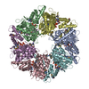
| ||||||||||||||||||||
|---|---|---|---|---|---|---|---|---|---|---|---|---|---|---|---|---|---|---|---|---|---|
| 1 |
| ||||||||||||||||||||
| Unit cell |
| ||||||||||||||||||||
| Noncrystallographic symmetry (NCS) | NCS oper:
|
-Components
| #1: Protein | / RUVBL1 / 49 KDA TATA BOX-BINDING PROTEIN-INTERACTING PROTEIN / 49 KDA TBP-INTERACTING PROTEIN / ...RUVBL1 / 49 KDA TATA BOX-BINDING PROTEIN-INTERACTING PROTEIN / 49 KDA TBP-INTERACTING PROTEIN / TIP49A / PONTIN 52 / NUCLEAR MATRIX PROTEIN 238 / NMP 238 / 54 KDA ERYTHROCYTE CYTOSOLIC PROTEIN / ECP-54 / TIP60-ASSOCIATED PROTEIN 54-ALPHA / TAP54-ALPHA / INO80 COMPLEX SUBUNIT H Mass: 40944.566 Da / Num. of mol.: 3 Source method: isolated from a genetically manipulated source Details: DOMAIN II WAS TRUNCATED BETWEEN RESIDUES T127 AND E233. A 4-RESIDUE LINKER WITH SEQUENCE GPPG WAS INSERTED TO REPLACE THE DELETED REGION Source: (gene. exp.) HOMO SAPIENS (human) / Plasmid: PETDUET / Production host:  ESCHERICHIA COLI BL21(DE3) (bacteria) / References: UniProt: Q9Y265, DNA helicase ESCHERICHIA COLI BL21(DE3) (bacteria) / References: UniProt: Q9Y265, DNA helicase#2: Protein | Mass: 42474.375 Da / Num. of mol.: 3 Source method: isolated from a genetically manipulated source Details: DOMAIN II WAS TRUNCATED BETWEEN RESIDUES E134 AND E237. A 4-RESIDUE LINKER WITH SEQUENCE GPPG WAS INSERTED TO REPLACE THE DELETED REGION Source: (gene. exp.) HOMO SAPIENS (human) / Plasmid: PETDUET / Production host: ESCHERICHIA COLI BL21(DE3) (bacteria) / References: UniProt: Q9Y230, DNA helicase#3: Chemical | ChemComp-ATP / Adenosine triphosphate  Mass: 507.181 Da / Num. of mol.: 6 / Source method: obtained synthetically / Formula: C10H16N5O13P3 / Comment: ATP, energy-carrying molecule*YM Mass: 507.181 Da / Num. of mol.: 6 / Source method: obtained synthetically / Formula: C10H16N5O13P3 / Comment: ATP, energy-carrying molecule*YMSequence details | CHAINS A-C TRUNCATED BETWEEN RESIDUES T127 AND E233 CHAINS D-F TRUNCATED BETWEEN RESIDUES E134 AND ...CHAINS A-C TRUNCATED BETWEEN RESIDUES T127 AND E233 CHAINS D-F TRUNCATED BETWEEN RESIDUES E134 AND E237 IN EACH CASE A 4-RESIDUE LINKER WITH SEQUENCE GPPG WAS INSERTED. | |
|---|
-Experimental details
-Experiment
| Experiment | Method: X-RAY DIFFRACTION / Number of used crystals: 1 |
|---|
- Sample preparation
| Crystal | Density Matthews: 2.6 Å3/Da / Density % sol: 53 % Description: THE SE ATOM SUBSTRUCTURE WAS DETERMINED BY MOLECULAR REPLACEMENT. |
|---|---|
| Crystal grow | pH: 7.5 Details: CRYSTALLIZATION DROPS WERE MIXED FROM EQUAL VOLUMES OF PROTEIN SOLUTION (12 MG/ML, 20MM TRIS-HCL PH 8.0, 200MM NACL, 10% GLYCEROL, 4MM MGCL2, 4MM ADP, 0.5MM TCEP) AND CRYSTALLIZATION ...Details: CRYSTALLIZATION DROPS WERE MIXED FROM EQUAL VOLUMES OF PROTEIN SOLUTION (12 MG/ML, 20MM TRIS-HCL PH 8.0, 200MM NACL, 10% GLYCEROL, 4MM MGCL2, 4MM ADP, 0.5MM TCEP) AND CRYSTALLIZATION SOLUTION (0.2M MGCL2, 30% PEG 400, 0.1M HEPES PH 7.5). |
-Data collection
| Diffraction | Mean temperature: 100 K |
|---|---|
| Diffraction source | Source: SYNCHROTRON / Site: ESRF  / Beamline: ID29 / Wavelength: 0.97633 / Beamline: ID29 / Wavelength: 0.97633 |
| Detector | Type: ADSC CCD / Detector: CCD / Date: Feb 12, 2010 / Details: MIRRORS |
| Radiation | Monochromator: SI(111) / Protocol: MAD / Monochromatic (M) / Laue (L): M / Scattering type: x-ray |
| Radiation wavelength | Wavelength: 0.97633 Å / Relative weight: 1 |
| Reflection | Resolution: 3→49 Å / Num. obs: 51774 / % possible obs: 99.7 % / Observed criterion σ(I): 0 / Redundancy: 4.2 % / Biso Wilson estimate: 99.32 Å2 / Rmerge(I) obs: 0.08 / Net I/σ(I): 12 |
| Reflection shell | Resolution: 3→3.16 Å / Redundancy: 4.4 % / Rmerge(I) obs: 0.76 / Mean I/σ(I) obs: 1.7 / % possible all: 100 |
- Processing
| Software |
| ||||||||||||||||||||||||||||||||||||||||||||||||||||||||||||||||||||||||||||||||||||||||||||||||||||||||||||||||||||||||||||||||||||||||||||||||||||||||||||||||||||||||||||||||||||||||||||||||||||||||||||||||||||||||||||||||||||||||||||||||||||||||||||||||||||||||||||||||||||||||||||||||||||||||||||||||||||||||||||||||||||||||||||||||||
|---|---|---|---|---|---|---|---|---|---|---|---|---|---|---|---|---|---|---|---|---|---|---|---|---|---|---|---|---|---|---|---|---|---|---|---|---|---|---|---|---|---|---|---|---|---|---|---|---|---|---|---|---|---|---|---|---|---|---|---|---|---|---|---|---|---|---|---|---|---|---|---|---|---|---|---|---|---|---|---|---|---|---|---|---|---|---|---|---|---|---|---|---|---|---|---|---|---|---|---|---|---|---|---|---|---|---|---|---|---|---|---|---|---|---|---|---|---|---|---|---|---|---|---|---|---|---|---|---|---|---|---|---|---|---|---|---|---|---|---|---|---|---|---|---|---|---|---|---|---|---|---|---|---|---|---|---|---|---|---|---|---|---|---|---|---|---|---|---|---|---|---|---|---|---|---|---|---|---|---|---|---|---|---|---|---|---|---|---|---|---|---|---|---|---|---|---|---|---|---|---|---|---|---|---|---|---|---|---|---|---|---|---|---|---|---|---|---|---|---|---|---|---|---|---|---|---|---|---|---|---|---|---|---|---|---|---|---|---|---|---|---|---|---|---|---|---|---|---|---|---|---|---|---|---|---|---|---|---|---|---|---|---|---|---|---|---|---|---|---|---|---|---|---|---|---|---|---|---|---|---|---|---|---|---|---|---|---|---|---|---|---|---|---|---|---|---|---|---|---|---|---|---|---|---|---|---|---|---|---|---|---|---|---|---|---|---|---|---|---|---|---|---|---|---|---|---|---|---|---|---|---|---|---|---|---|---|---|---|---|
| Refinement | Method to determine structure: MAD Starting model: PDB ENTRY 2C9O Resolution: 3→46.14 Å / Cor.coef. Fo:Fc: 0.9462 / Cor.coef. Fo:Fc free: 0.9413 / Cross valid method: THROUGHOUT / σ(F): 0 / SU Rfree Blow DPI: 0.316
| ||||||||||||||||||||||||||||||||||||||||||||||||||||||||||||||||||||||||||||||||||||||||||||||||||||||||||||||||||||||||||||||||||||||||||||||||||||||||||||||||||||||||||||||||||||||||||||||||||||||||||||||||||||||||||||||||||||||||||||||||||||||||||||||||||||||||||||||||||||||||||||||||||||||||||||||||||||||||||||||||||||||||||||||||||
| Displacement parameters | Biso mean: 111.89 Å2
| ||||||||||||||||||||||||||||||||||||||||||||||||||||||||||||||||||||||||||||||||||||||||||||||||||||||||||||||||||||||||||||||||||||||||||||||||||||||||||||||||||||||||||||||||||||||||||||||||||||||||||||||||||||||||||||||||||||||||||||||||||||||||||||||||||||||||||||||||||||||||||||||||||||||||||||||||||||||||||||||||||||||||||||||||||
| Refine analyze | Luzzati coordinate error obs: 0.577 Å | ||||||||||||||||||||||||||||||||||||||||||||||||||||||||||||||||||||||||||||||||||||||||||||||||||||||||||||||||||||||||||||||||||||||||||||||||||||||||||||||||||||||||||||||||||||||||||||||||||||||||||||||||||||||||||||||||||||||||||||||||||||||||||||||||||||||||||||||||||||||||||||||||||||||||||||||||||||||||||||||||||||||||||||||||||
| Refinement step | Cycle: LAST / Resolution: 3→46.14 Å
| ||||||||||||||||||||||||||||||||||||||||||||||||||||||||||||||||||||||||||||||||||||||||||||||||||||||||||||||||||||||||||||||||||||||||||||||||||||||||||||||||||||||||||||||||||||||||||||||||||||||||||||||||||||||||||||||||||||||||||||||||||||||||||||||||||||||||||||||||||||||||||||||||||||||||||||||||||||||||||||||||||||||||||||||||||
| Refine LS restraints |
| ||||||||||||||||||||||||||||||||||||||||||||||||||||||||||||||||||||||||||||||||||||||||||||||||||||||||||||||||||||||||||||||||||||||||||||||||||||||||||||||||||||||||||||||||||||||||||||||||||||||||||||||||||||||||||||||||||||||||||||||||||||||||||||||||||||||||||||||||||||||||||||||||||||||||||||||||||||||||||||||||||||||||||||||||||
| LS refinement shell | Resolution: 3→3.08 Å / Total num. of bins used: 20
| ||||||||||||||||||||||||||||||||||||||||||||||||||||||||||||||||||||||||||||||||||||||||||||||||||||||||||||||||||||||||||||||||||||||||||||||||||||||||||||||||||||||||||||||||||||||||||||||||||||||||||||||||||||||||||||||||||||||||||||||||||||||||||||||||||||||||||||||||||||||||||||||||||||||||||||||||||||||||||||||||||||||||||||||||||
| Refinement TLS params. | Refine-ID: X-RAY DIFFRACTION
| ||||||||||||||||||||||||||||||||||||||||||||||||||||||||||||||||||||||||||||||||||||||||||||||||||||||||||||||||||||||||||||||||||||||||||||||||||||||||||||||||||||||||||||||||||||||||||||||||||||||||||||||||||||||||||||||||||||||||||||||||||||||||||||||||||||||||||||||||||||||||||||||||||||||||||||||||||||||||||||||||||||||||||||||||||
| Refinement TLS group |
|
