 Movie
Movie Controller


+ Open data

- Basic information
| Entry | Database: PDB / ID: 2xol | ||||||
|---|---|---|---|---|---|---|---|
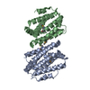
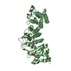
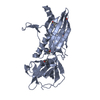
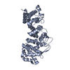
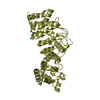
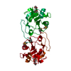
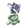
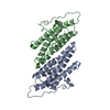
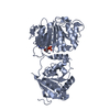
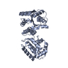
| Title | High resolution structure of TtrD from Archaeoglobus fulgidus | ||||||
 Components Components | CHAPERONE PROTEIN TTRD | ||||||
 Keywords Keywords | CHAPERONE / TAT SYSTEM | ||||||
| Function / homology | TorD-like / TorD-like / DMSO/Nitrate reductase chaperone / TorD-like superfamily / Nitrate reductase delta subunit / Orthogonal Bundle / Mainly Alpha / cytoplasm / Tat proofreading chaperone TtrD Function and homology information Function and homology information | ||||||
| Biological species |   ARCHAEOGLOBUS FULGIDUS (archaea) ARCHAEOGLOBUS FULGIDUS (archaea) | ||||||
| Method | X-RAY DIFFRACTION / SYNCHROTRON / MOLECULAR REPLACEMENT / Resolution: 1.35 Å | ||||||
 Authors Authors | Dawson, A. / Coulthurst, S.J. / Sargent, F. / Hunter, W.N. | ||||||
 Citation Citation | Journal: Biochemistry / Year: 2012 Title: Conserved Signal Peptide Recognition Systems Across the Prokaryotic Domains. Authors: Coulthurst, S.J. / Dawson, A. / Hunter, W.N. / Sargent, F. | ||||||
| History |
|
- Structure visualization
| Structure viewer | Molecule: MolmilJmol/JSmol |
|---|
- Downloads & links
-Download
| PDBx/mmCIF format | 2xol.cif.gz | 168.7 KB | Display | PDBx/mmCIF format |
|---|---|---|---|---|
| PDB format | pdb2xol.ent.gz | 135.1 KB | Display | PDB format |
| PDBx/mmJSON format | 2xol.json.gz | Tree view | PDBx/mmJSON format | |
| Others |  Other downloads Other downloads |
-Validation report
| Arichive directory | https://data.pdbj.org/pub/pdb/validation_reports/xo/2xolftp://data.pdbj.org/pub/pdb/validation_reports/xo/2xol | HTTPS FTP |
|---|
-Related structure data
| Related structure data |  2y6yC  2yjmC  2idgS C: citing same article ( S: Starting model for refinement |
|---|---|
| Similar structure data |
-Links
 PDBj
PDBj
- Assembly
| Deposited unit | 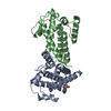
| ||||||||
|---|---|---|---|---|---|---|---|---|---|
| 1 |
| ||||||||
| Unit cell |
|
-Components
| #1: Protein | Mass: 20396.436 Da / Num. of mol.: 2 Source method: isolated from a genetically manipulated source Source: (gene. exp.) ARCHAEOGLOBUS FULGIDUS (archaea) / Plasmid: PET15BTEV_TTRD / Production host:  ESCHERICHIA COLI (E. coli) / Strain (production host): BL21(DE3) / References: UniProt: O30077 ESCHERICHIA COLI (E. coli) / Strain (production host): BL21(DE3) / References: UniProt: O30077#2: Chemical | ChemComp-EDO / | Ethylene glycol  Mass: 62.068 Da / Num. of mol.: 1 / Source method: obtained synthetically / Formula: C2H6O2 Mass: 62.068 Da / Num. of mol.: 1 / Source method: obtained synthetically / Formula: C2H6O2#3: Water | ChemComp-HOH / | Water Mass: 18.015 Da / Num. of mol.: 393 / Source method: isolated from a natural source / Formula: H2O Mass: 18.015 Da / Num. of mol.: 393 / Source method: isolated from a natural source / Formula: H2OSequence details | ADDITIONAL | |
|---|
-Experimental details
-Experiment
| Experiment | Method: X-RAY DIFFRACTION / Number of used crystals: 1 |
|---|
- Sample preparation
| Crystal | Density Matthews: 2.63 Å3/Da / Density % sol: 53.2 % / Description: NONE |
|---|---|
| Crystal grow | pH: 7.5 / Details: 0.2 M SODIUM FORMATE, 20% PEG 3350, pH 7.5 |
-Data collection
| Diffraction | Mean temperature: 100 K |
|---|---|
| Diffraction source | Source: SYNCHROTRON / Site: ESRF  / Beamline: ID29 / Wavelength: 0.977 / Beamline: ID29 / Wavelength: 0.977 |
| Detector | Type: ADSC CCD / Detector: CCD / Date: Feb 14, 2010 |
| Radiation | Protocol: SINGLE WAVELENGTH / Monochromatic (M) / Laue (L): M / Scattering type: x-ray |
| Radiation wavelength | Wavelength: 0.977 Å / Relative weight: 1 |
| Reflection | Resolution: 1.35→64.69 Å / Num. obs: 89700 / % possible obs: 96.9 % / Observed criterion σ(I): 0 / Redundancy: 6.9 % / Biso Wilson estimate: 14.78 Å2 / Rmerge(I) obs: 0.05 / Net I/σ(I): 17.6 |
| Reflection shell | Resolution: 1.35→1.42 Å / Redundancy: 6.3 % / Rmerge(I) obs: 0.49 / Mean I/σ(I) obs: 4.2 / % possible all: 94.9 |
- Processing
| Software |
| ||||||||||||||||||||||||||||||||||||||||||||||||||||||||||||||||||||||||||||||||||||||||||||||||||||||||||||||||||||||||||||||||||||||||||||||||||||||||||||||||||||||||||||||||||||||
|---|---|---|---|---|---|---|---|---|---|---|---|---|---|---|---|---|---|---|---|---|---|---|---|---|---|---|---|---|---|---|---|---|---|---|---|---|---|---|---|---|---|---|---|---|---|---|---|---|---|---|---|---|---|---|---|---|---|---|---|---|---|---|---|---|---|---|---|---|---|---|---|---|---|---|---|---|---|---|---|---|---|---|---|---|---|---|---|---|---|---|---|---|---|---|---|---|---|---|---|---|---|---|---|---|---|---|---|---|---|---|---|---|---|---|---|---|---|---|---|---|---|---|---|---|---|---|---|---|---|---|---|---|---|---|---|---|---|---|---|---|---|---|---|---|---|---|---|---|---|---|---|---|---|---|---|---|---|---|---|---|---|---|---|---|---|---|---|---|---|---|---|---|---|---|---|---|---|---|---|---|---|---|---|
| Refinement | Method to determine structure: MOLECULAR REPLACEMENT Starting model: PDB ENTRY 2IDG Resolution: 1.35→64.93 Å / Cor.coef. Fo:Fc: 0.971 / Cor.coef. Fo:Fc free: 0.963 / SU B: 1.499 / SU ML: 0.028 / Cross valid method: THROUGHOUT / ESU R: 0.051 / ESU R Free: 0.048 / Stereochemistry target values: MAXIMUM LIKELIHOOD Details: HYDROGENS HAVE BEEN ADDED IN THE RIDING POSITIONS. U VALUES REFINED INDIVIDUALLY.
| ||||||||||||||||||||||||||||||||||||||||||||||||||||||||||||||||||||||||||||||||||||||||||||||||||||||||||||||||||||||||||||||||||||||||||||||||||||||||||||||||||||||||||||||||||||||
| Solvent computation | Ion probe radii: 0.8 Å / Shrinkage radii: 0.8 Å / VDW probe radii: 1.4 Å / Solvent model: MASK | ||||||||||||||||||||||||||||||||||||||||||||||||||||||||||||||||||||||||||||||||||||||||||||||||||||||||||||||||||||||||||||||||||||||||||||||||||||||||||||||||||||||||||||||||||||||
| Displacement parameters | Biso mean: 16.751 Å2
| ||||||||||||||||||||||||||||||||||||||||||||||||||||||||||||||||||||||||||||||||||||||||||||||||||||||||||||||||||||||||||||||||||||||||||||||||||||||||||||||||||||||||||||||||||||||
| Refinement step | Cycle: LAST / Resolution: 1.35→64.93 Å
| ||||||||||||||||||||||||||||||||||||||||||||||||||||||||||||||||||||||||||||||||||||||||||||||||||||||||||||||||||||||||||||||||||||||||||||||||||||||||||||||||||||||||||||||||||||||
| Refine LS restraints |
|