[English] 日本語
Yorodumi
- PDB-2xkj: CRYSTAL STRUCTURE OF CATALYTIC CORE OF A. BAUMANNII TOPO IV (PARE... -

+
Open data


ID or keywords:

Loading...

-
Basic information

Entry
Database: PDB / ID: 2xkj
Title
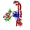
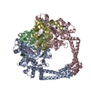
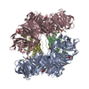
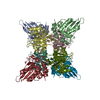
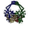
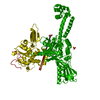
CRYSTAL STRUCTURE OF CATALYTIC CORE OF A. BAUMANNII TOPO IV (PARE- PARC FUSION TRUNCATE)
ComponentsTOPOISOMERASE IV
KeywordsISOMERASE / TYPE IIA TOPOISOMERASE
Function / homology
Function and homology information


DNA topoisomerase type II (double strand cut, ATP-hydrolyzing) activity / DNA topoisomerase (ATP-hydrolysing) / extrinsic component of plasma membrane / DNA topological change / chromosome segregation / chromosome / DNA binding / ATP binding / cytoplasm
Similarity search - Function
DNA topoisomerase IV, subunit A, Gram-negative / DNA topoisomerase IV, subunit B, Gram-negative / Rossmann fold - #670 / DNA gyrase/topoisomerase IV, subunit A, C-terminal repeat / DNA gyrase/topoisomerase IV, subunit A, C-terminal / DNA gyrase C-terminal domain, beta-propeller / DNA topoisomerase, type IIA, alpha-helical domain superfamily / DNA topoisomerase, type IIA, domain A / DNA topoisomerase, type IIA, domain A, alpha-beta / DNA gyrase/topoisomerase IV, subunit A ...DNA topoisomerase IV, subunit A, Gram-negative / DNA topoisomerase IV, subunit B, Gram-negative / Rossmann fold - #670 / DNA gyrase/topoisomerase IV, subunit A, C-terminal repeat / DNA gyrase/topoisomerase IV, subunit A, C-terminal / DNA gyrase C-terminal domain, beta-propeller / DNA topoisomerase, type IIA, alpha-helical domain superfamily / DNA topoisomerase, type IIA, domain A / DNA topoisomerase, type IIA, domain A, alpha-beta / DNA gyrase/topoisomerase IV, subunit A / DNA Topoisomerase IV / DNA gyrase B subunit, C-terminal / DNA gyrase B subunit, carboxyl terminus / DNA topoisomerase, type IIA, subunit B, domain 2 / DNA gyrase B / DNA topoisomerase, type IIA / DNA topoisomerase, type IIA, conserved site / DNA topoisomerase II signature. / TopoisomeraseII / DNA topoisomerase, type IIA, subunit B, C-terminal / Toprim domain / DNA topoisomerase, type IIA-like domain superfamily / Toprim domain profile. / TOPRIM domain / Histidine kinase-, DNA gyrase B-, and HSP90-like ATPase / Histidine kinase-like ATPases / Histidine kinase/HSP90-like ATPase / Histidine kinase/HSP90-like ATPase superfamily / Ribosomal protein S5 domain 2-type fold, subgroup / Ribosomal protein S5 domain 2-type fold / Rossmann fold / 3-Layer(aba) Sandwich / Alpha Beta
Similarity search - Domain/homology
DNA topoisomerase 4 subunit B / DNA topoisomerase 4 subunit A
Similarity search - Component
Biological speciesACINETOBACTER BAUMANNII (bacteria)
MethodX-RAY DIFFRACTION / SYNCHROTRON / MOLECULAR REPLACEMENT / Resolution: 2.2 Å
AuthorsWohlkonig, A. / Chan, P.F. / Fosberry, A.P. / Homes, P. / Huang, J. / Kranz, M. / Leydon, V.R. / Miles, T.J. / Pearson, N.D. / Perera, R.L. ...Wohlkonig, A. / Chan, P.F. / Fosberry, A.P. / Homes, P. / Huang, J. / Kranz, M. / Leydon, V.R. / Miles, T.J. / Pearson, N.D. / Perera, R.L. / Shillings, A.J. / Gwynn, M.N. / Bax, B.D.
CitationJournal: Nat.Struct.Mol.Biol. / Year: 2010
Title: Structural Basis of Quinolone Inhibition of Type Iia Topoisomerases and Target-Mediated Resistance
Authors: Wohlkonig, A. / Chan, P.F. / Fosberry, A.P. / Homes, P. / Huang, J. / Kranz, M. / Leydon, V.R. / Miles, T.J. / Pearson, N.D. / Perera, R.L. / Shillings, A.J. / Gwynn, M.N. / Bax, B.D.
History
DepositionJul 8, 2010Deposition site: PDBE / Processing site: PDBE
Revision 1.0Sep 1, 2010Provider: repository / Type: Initial release
Revision 1.1May 19, 2011Group: Version format compliance
Revision 1.2Jul 13, 2011Group: Version format compliance
Revision 1.3May 8, 2019Group: Data collection / Experimental preparation / Other
Category: database_PDB_rev / database_PDB_rev_record ...database_PDB_rev / database_PDB_rev_record / exptl_crystal_grow / pdbx_database_proc / pdbx_database_status
Item: _exptl_crystal_grow.method / _pdbx_database_status.recvd_author_approval
Revision 1.4Sep 25, 2019Group: Data collection / Other / Category: pdbx_database_status / reflns
Item: _pdbx_database_status.status_code_sf / _reflns.pdbx_Rmerge_I_obs
Revision 1.5Dec 20, 2023Group: Data collection / Database references ...Data collection / Database references / Derived calculations / Refinement description
Category: chem_comp_atom / chem_comp_bond ...chem_comp_atom / chem_comp_bond / database_2 / pdbx_initial_refinement_model / struct_site
Item: _database_2.pdbx_DOI / _database_2.pdbx_database_accession ..._database_2.pdbx_DOI / _database_2.pdbx_database_accession / _struct_site.pdbx_auth_asym_id / _struct_site.pdbx_auth_comp_id / _struct_site.pdbx_auth_seq_id
Remark 650 HELIX DETERMINATION METHOD: AUTHOR PROVIDED.

-
Structure visualization

Structure viewerMolecule:
MolmilJmol/JSmol

Downloads & links

-
Assembly

Deposited unit
E: TOPOISOMERASE IV
hetero molecules


Theoretical massNumber of molelcules
Total (without water)86,3704
Polymers86,0861
Non-polymers2843
Water9,620534
1
E: TOPOISOMERASE IV
hetero molecules

E: TOPOISOMERASE IV
hetero molecules


Theoretical massNumber of molelcules
Total (without water)172,7408
Polymers172,1722
Non-polymers5686
Water362
TypeNameSymmetry operationNumber
identity operation1_555x,y,z1
crystal symmetry operation2_555-x,y,-z1
Buried area7170 Å2
ΔGint-38.2 kcal/mol
Surface area61590 Å2
MethodPISA
Unit cell
Length a, b, c (Å)163.389, 74.433, 80.211
Angle α, β, γ (deg.)90.00, 107.96, 90.00
Int Tables number5
Space group name H-MC121
Components on special symmetry positions
IDModelComponents
11E-3081-

HOH

DetailsDIMER SITS ON CRYSTALLOGRAPHIC TWO FOLD AXIS.

-
Components

#1: Protein TOPOISOMERASE IV /


Mass: 86085.773 Da / Num. of mol.: 1
Fragment: PARE SUBUNIT C-TERMINAL 28KDA DOMAIN, RESIDUES 370-627, PARC SUBUNIT N-TERMINAL 58KDA DOMAIN, RESIDUES 1 TO 503
Source method: isolated from a genetically manipulated source
Details: THE CONSTRUCT IS A FUSION OF THE TOPO IV, PARE AND PARC SUBUNITS. RESIDUE 604 OF PARE IS LINKED TO RESIDUES 996 OF PARC
Source: (gene. exp.) ACINETOBACTER BAUMANNII (bacteria) / Strain: EUROFINS MEDINET / Production host: ESCHERICHIA COLI (E. coli) / Strain (production host): BL21(DE3) / References: UniProt: B0V9T6, UniProt: B0VP98, EC: 5.99.1.-
#2: Chemical ChemComp-SO4 / SULFATE ION / Sulfate


Mass: 96.063 Da / Num. of mol.: 2 / Source method: obtained synthetically / Formula: SO4
#3: Chemical ChemComp-GOL / GLYCEROL / GLYCERIN / PROPANE-1,2,3-TRIOL / Glycerol


Mass: 92.094 Da / Num. of mol.: 1 / Source method: obtained synthetically / Formula: C3H8O3
#4: Water ChemComp-HOH / water / Water


Mass: 18.015 Da / Num. of mol.: 534 / Source method: isolated from a natural source / Formula: H2O

-
Experimental details

-
Experiment

ExperimentMethod: X-RAY DIFFRACTION / Number of used crystals: 1

-
Sample preparation

CrystalDensity Matthews: 2.69 Å3/Da / Density % sol: 54.36 % / Description: NONE
Crystal growMethod: vapor diffusion, hanging drop
Details: HANGING DROP VAPOUR DIFFUSION METHOD USING 0.2 M LITHIUM SULFATE, 0.1 M TRIS PH 8.5, 16% PEG 4000 AS THE PRECIPITATING AGENT

-
Data collection

DiffractionMean temperature: 100 K
Diffraction sourceSource: SYNCHROTRON / Site: ESRF / Beamline: ID14-4 / Wavelength: 0.9755
DetectorType: ADSC CCD / Detector: CCD
RadiationProtocol: SINGLE WAVELENGTH / Monochromatic (M) / Laue (L): M / Scattering type: x-ray
Radiation wavelengthWavelength: 0.9755 Å / Relative weight: 1
ReflectionResolution: 2.2→40 Å / Num. obs: 46394 / % possible obs: 99.9 % / Observed criterion σ(I): 0 / Redundancy: 3.7 % / Biso Wilson estimate: 26.56 Å2 / Rmerge(I) obs: 0.087 / Net I/σ(I): 13.7
Reflection shellResolution: 2.2→2.24 Å / Redundancy: 3.7 % / Rmerge(I) obs: 0.41 / Mean I/σ(I) obs: 3.5 / % possible all: 100

-
Processing

Software
NameVersionClassification
PHENIX(PHENIX.REFINE)refinement
HKLdata reduction
SCALEPACKdata scaling
PHASERphasing
RefinementMethod to determine structure: MOLECULAR REPLACEMENT
Starting model: DOMAINS FROM PDB ENTRY 2XCS
Resolution: 2.2→24.909 Å / SU ML: 0.27 / σ(F): 1.34 / Phase error: 20.69 / Stereochemistry target values: ML
RfactorNum. reflection% reflection
Rfree0.2129 2342 5.1 %
Rwork0.1608 --
obs0.1634 46360 99.75 %
Solvent computationShrinkage radii: 0.9 Å / VDW probe radii: 1.11 Å / Solvent model: FLAT BULK SOLVENT MODEL / Bsol: 47.337 Å2 / ksol: 0.331 e/Å3
Displacement parametersBiso mean: 30.3 Å2
Baniso -1Baniso -2Baniso -3
1--2.2682 Å20 Å2-0.8283 Å2
2--0.9214 Å20 Å2
3---1.3469 Å2
Refinement stepCycle: LAST / Resolution: 2.2→24.909 Å
ProteinNucleic acidLigandSolventTotal
Num. atoms5628 0 16 534 6178
Refine LS restraints
Refine-IDTypeDev idealNumber
X-RAY DIFFRACTIONf_bond_d0.0085896
X-RAY DIFFRACTIONf_angle_d1.098002
X-RAY DIFFRACTIONf_dihedral_angle_d17.5722252
X-RAY DIFFRACTIONf_chiral_restr0.07908
X-RAY DIFFRACTIONf_plane_restr0.0041054
LS refinement shell
Resolution (Å)Rfactor RfreeNum. reflection RfreeRfactor RworkNum. reflection RworkRefine-ID% reflection obs (%)
2.2027-2.24760.28721140.18912562X-RAY DIFFRACTION98
2.2476-2.29650.28271320.18822592X-RAY DIFFRACTION100
2.2965-2.34990.25711530.18232556X-RAY DIFFRACTION100
2.3499-2.40860.26081170.18172573X-RAY DIFFRACTION100
2.4086-2.47360.23991370.17692597X-RAY DIFFRACTION100
2.4736-2.54640.24271500.16562566X-RAY DIFFRACTION100
2.5464-2.62850.22761640.17352545X-RAY DIFFRACTION100
2.6285-2.72230.24421190.16722621X-RAY DIFFRACTION100
2.7223-2.83110.20731470.16522559X-RAY DIFFRACTION100
2.8311-2.95980.2171270.16222614X-RAY DIFFRACTION100
2.9598-3.11560.23261400.16872578X-RAY DIFFRACTION100
3.1156-3.31040.21061440.16722589X-RAY DIFFRACTION100
3.3104-3.56530.21511490.15732572X-RAY DIFFRACTION100
3.5653-3.92280.1841460.14392610X-RAY DIFFRACTION100
3.9228-4.48760.1521350.12682588X-RAY DIFFRACTION100
4.4876-5.6430.1911260.13832636X-RAY DIFFRACTION100
5.643-24.91060.18341420.15562660X-RAY DIFFRACTION100

+
About Yorodumi

-
News

-
Feb 9, 2022. New format data for meta-information of EMDB entries

New format data for meta-information of EMDB entries

  • Version 3 of the EMDB header file is now the official format.
  • The previous official version 1.9 will be removed from the archive.

Related info.:EMDB header

External links:wwPDB to switch to version 3 of the EMDB data model

-
Aug 12, 2020. Covid-19 info

Covid-19 info

URL: https://pdbj.org/emnavi/covid19.php

New page: Covid-19 featured information page in EM Navigator.

Related info.:Covid-19 info / Mar 5, 2020. Novel coronavirus structure data

+
Mar 5, 2020. Novel coronavirus structure data

Novel coronavirus structure data

Related info.:Yorodumi Speices / Aug 12, 2020. Covid-19 info

External links:COVID-19 featured content - PDBj / Molecule of the Month (242):Coronavirus Proteases

+
Jan 31, 2019. EMDB accession codes are about to change! (news from PDBe EMDB page)

EMDB accession codes are about to change! (news from PDBe EMDB page)

  • The allocation of 4 digits for EMDB accession codes will soon come to an end. Whilst these codes will remain in use, new EMDB accession codes will include an additional digit and will expand incrementally as the available range of codes is exhausted. The current 4-digit format prefixed with “EMD-” (i.e. EMD-XXXX) will advance to a 5-digit format (i.e. EMD-XXXXX), and so on. It is currently estimated that the 4-digit codes will be depleted around Spring 2019, at which point the 5-digit format will come into force.
  • The EM Navigator/Yorodumi systems omit the EMD- prefix.

Related info.:Q: What is EMD? / ID/Accession-code notation in Yorodumi/EM Navigator

External links:EMDB Accession Codes are Changing Soon! / Contact to PDBj

+
Jul 12, 2017. Major update of PDB

Major update of PDB

  • wwPDB released updated PDB data conforming to the new PDBx/mmCIF dictionary.
  • This is a major update changing the version number from 4 to 5, and with Remediation, in which all the entries are updated.
  • In this update, many items about electron microscopy experimental information are reorganized (e.g. em_software).
  • Now, EM Navigator and Yorodumi are based on the updated data.

External links:wwPDB Remediation / Enriched Model Files Conforming to OneDep Data Standards Now Available in the PDB FTP Archive

-
Yorodumi

Thousand views of thousand structures

  • Yorodumi is a browser for structure data from EMDB, PDB, SASBDB, etc.
  • This page is also the successor to EM Navigator detail page, and also detail information page/front-end page for Omokage search.
  • The word "yorodu" (or yorozu) is an old Japanese word meaning "ten thousand". "mi" (miru) is to see.

Related info.:EMDB / PDB / SASBDB / Comparison of 3 databanks / Yorodumi Search / Aug 31, 2016. New EM Navigator & Yorodumi / Yorodumi Papers / Jmol/JSmol / Function and homology information / Changes in new EM Navigator and Yorodumi

Read more