 Movie
Movie Controller

[English] 日本語
 Yorodumi
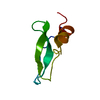
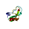
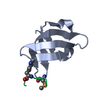
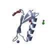
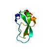
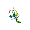
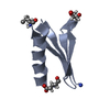
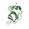
- PDB-2x9b: The filamentous phages fd and IF1 use different infection mechanisms -
+ Open data

- Basic information
| Entry | Database: PDB / ID: 2x9b | ||||||
|---|---|---|---|---|---|---|---|
| Title | The filamentous phages fd and IF1 use different infection mechanisms | ||||||
 Components Components | ATTACHMENT PROTEIN G3P | ||||||
 Keywords Keywords |  VIRAL PROTEIN / VIRION / PHAGE RECOGNITION / HOST PHAGE COAT PROTEIN HOST-VIRUS INTERACTION VIRAL PROTEIN / VIRION / PHAGE RECOGNITION / HOST PHAGE COAT PROTEIN HOST-VIRUS INTERACTION | ||||||
| Function / homology |  Function and homology information Function and homology information: / viral extrusion / virion attachment to host cell pilus / adhesion receptor-mediated virion attachment to host cell / host cell membrane / viral capsid / entry receptor-mediated virion attachment to host cell / membraneSimilarity search - Function | ||||||
| Biological species |  ENTEROBACTERIA PHAGE IF1 (virus) ENTEROBACTERIA PHAGE IF1 (virus) | ||||||
| Method | X-RAY DIFFRACTION / SYNCHROTRON / MOLECULAR REPLACEMENT / Resolution: 2.92 Å | ||||||
 Authors Authors | Lorenz, S.H. / Jakob, R.P. / Weininger, U. / Dobbek, H. / Schmid, F.X. | ||||||
 Citation Citation | Journal: J.Mol.Biol. / Year: 2011 Title: The Filamentous Phages Fd and If1 Use Different Mechanisms to Infect Escherichia Coli. Authors: Lorenz, S.H. / Jakob, R.P. / Weininger, U. / Balbach, J. / Dobbek, H. / Schmid, F.X. | ||||||
| History |
|
- Structure visualization
| Structure viewer | Molecule: MolmilJmol/JSmol |
|---|
- Downloads & links
-Download
| PDBx/mmCIF format | 2x9b.cif.gz | 60.7 KB | Display | PDBx/mmCIF format |
|---|---|---|---|---|
| PDB format | pdb2x9b.ent.gz | 46 KB | Display | PDB format |
| PDBx/mmJSON format | 2x9b.json.gz | Tree view | PDBx/mmJSON format | |
| Others |  Other downloads Other downloads |
-Validation report
| Arichive directory | https://data.pdbj.org/pub/pdb/validation_reports/x9/2x9bftp://data.pdbj.org/pub/pdb/validation_reports/x9/2x9b | HTTPS FTP |
|---|
-Related structure data
| Related structure data |  2x9aC 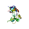 1g3pS C: citing same article ( S: Starting model for refinement |
|---|---|
| Similar structure data |

-Links
 PDBj
PDBj- Assembly
| Deposited unit | 
| ||||||||
|---|---|---|---|---|---|---|---|---|---|
| 1 | 
| ||||||||
| 2 | 
| ||||||||
| Unit cell |
|
-Components
| #1: Protein | Mass: 7037.493 Da / Num. of mol.: 2 / Fragment: TOLA-BINDING DOMAIN, RESIDUES 17-81 Source method: isolated from a genetically manipulated source Source: (gene. exp.) ENTEROBACTERIA PHAGE IF1 (virus) / Plasmid: PET11A / Production host:  ESCHERICHIA COLI (E. coli) / Strain (production host): BL21(DE3) / References: UniProt: O80297 ESCHERICHIA COLI (E. coli) / Strain (production host): BL21(DE3) / References: UniProt: O80297#2: Water | ChemComp-HOH / | Water Mass: 18.015 Da / Num. of mol.: 48 / Source method: isolated from a natural source / Formula: H2O Mass: 18.015 Da / Num. of mol.: 48 / Source method: isolated from a natural source / Formula: H2O |
|---|
-Experimental details
-Experiment
| Experiment | Method: X-RAY DIFFRACTION / Number of used crystals: 1 |
|---|
- Sample preparation
| Crystal | Density Matthews: 4.42 Å3/Da / Density % sol: 71.97 % / Description: NONE |
|---|---|
| Crystal grow | pH: 6.5 Details: 3 M NACL, 5 % MPD, 0.1 M CACL2, 0.1 M IMIDAZOL PH 6.5 |
-Data collection
| Diffraction | Mean temperature: 100 K |
|---|---|
| Diffraction source | Source: SYNCHROTRON / Site: BESSY  / Beamline: 14.1 / Wavelength: 0.914 / Beamline: 14.1 / Wavelength: 0.914 |
| Detector | Type: MARRESEARCH / Detector: CCD |
| Radiation | Protocol: SINGLE WAVELENGTH / Monochromatic (M) / Laue (L): M / Scattering type: x-ray |
| Radiation wavelength | Wavelength: 0.914 Å / Relative weight: 1 |
| Reflection | Resolution: 2.92→31.72 Å / Num. obs: 5889 / % possible obs: 99.7 % / Observed criterion σ(I): 3.5 / Redundancy: 11.38 % / Biso Wilson estimate: 71.67 Å2 / Rmerge(I) obs: 0.1 / Net I/σ(I): 20.6 |
| Reflection shell | Resolution: 2.92→3.26 Å / Redundancy: 11.8 % / Rmerge(I) obs: 0.68 / Mean I/σ(I) obs: 3.87 / % possible all: 100 |
- Processing
| Software |
| ||||||||||||||||||||||||||||||||||||||||||||||||||||||||||||||||||||||||||||||||||||||||||||||||||||||||||||||||||
|---|---|---|---|---|---|---|---|---|---|---|---|---|---|---|---|---|---|---|---|---|---|---|---|---|---|---|---|---|---|---|---|---|---|---|---|---|---|---|---|---|---|---|---|---|---|---|---|---|---|---|---|---|---|---|---|---|---|---|---|---|---|---|---|---|---|---|---|---|---|---|---|---|---|---|---|---|---|---|---|---|---|---|---|---|---|---|---|---|---|---|---|---|---|---|---|---|---|---|---|---|---|---|---|---|---|---|---|---|---|---|---|---|---|---|---|
| Refinement | Method to determine structure: MOLECULAR REPLACEMENT Starting model: PDB ENTRY 1G3P Resolution: 2.92→31.72 Å / Cor.coef. Fo:Fc: 0.8726 / Cor.coef. Fo:Fc free: 0.8215 / Cross valid method: THROUGHOUT / σ(F): 0
| ||||||||||||||||||||||||||||||||||||||||||||||||||||||||||||||||||||||||||||||||||||||||||||||||||||||||||||||||||
| Displacement parameters | Biso mean: 54.83 Å2
| ||||||||||||||||||||||||||||||||||||||||||||||||||||||||||||||||||||||||||||||||||||||||||||||||||||||||||||||||||
| Refine analyze | Luzzati coordinate error obs: 0.452 Å | ||||||||||||||||||||||||||||||||||||||||||||||||||||||||||||||||||||||||||||||||||||||||||||||||||||||||||||||||||
| Refinement step | Cycle: LAST / Resolution: 2.92→31.72 Å
| ||||||||||||||||||||||||||||||||||||||||||||||||||||||||||||||||||||||||||||||||||||||||||||||||||||||||||||||||||
| Refine LS restraints |
| ||||||||||||||||||||||||||||||||||||||||||||||||||||||||||||||||||||||||||||||||||||||||||||||||||||||||||||||||||
| LS refinement shell | Resolution: 2.92→3.26 Å / Total num. of bins used: 5
| ||||||||||||||||||||||||||||||||||||||||||||||||||||||||||||||||||||||||||||||||||||||||||||||||||||||||||||||||||
| Refinement TLS params. | Method: refined / Refine-ID: X-RAY DIFFRACTION
| ||||||||||||||||||||||||||||||||||||||||||||||||||||||||||||||||||||||||||||||||||||||||||||||||||||||||||||||||||
| Refinement TLS group |
|
