 Movie
Movie Controller

[English] 日本語
 Yorodumi
- PDB-2x8g: Oxidized thioredoxin glutathione reductase from Schistosoma mansoni -
+ Open data

- Basic information
| Entry | Database: PDB / ID: 2x8g | ||||||
|---|---|---|---|---|---|---|---|
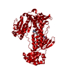
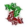
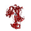
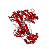
| Title | Oxidized thioredoxin glutathione reductase from Schistosoma mansoni | ||||||
 Components Components | THIOREDOXIN GLUTATHIONE REDUCTASE | ||||||
 Keywords Keywords |  OXIDOREDUCTASE / REDOX-ACTIVE CENTER / DETOXIFICATION PATHWAY / FLAVOPROTEIN OXIDOREDUCTASE / REDOX-ACTIVE CENTER / DETOXIFICATION PATHWAY / FLAVOPROTEIN | ||||||
| Function / homology |  Function and homology informationthioredoxin-disulfide reductase / thioredoxin-disulfide reductase (NADPH) activity / cell redox homeostasis / flavin adenine dinucleotide binding / metal ion binding Function and homology informationthioredoxin-disulfide reductase / thioredoxin-disulfide reductase (NADPH) activity / cell redox homeostasis / flavin adenine dinucleotide binding / metal ion bindingSimilarity search - Function | ||||||
| Biological species |  SCHISTOSOMA MANSONI (invertebrata) SCHISTOSOMA MANSONI (invertebrata) | ||||||
| Method | X-RAY DIFFRACTION / SYNCHROTRON / MOLECULAR REPLACEMENT / Resolution: 1.9 Å | ||||||
 Authors Authors | Angelucci, F. / Dimastrogiovanni, D. / Boumis, G. / Brunori, M. / Miele, A.E. / Saccoccia, F. / Bellelli, A. | ||||||
 Citation Citation | Journal: J.Biol.Chem. / Year: 2010 Title: Mapping the Catalytic Cycle of Schistosoma Mansoni Thioredoxin Glutathione Reductase by X-Ray Crystallography Authors: Angelucci, F. / Dimastrogiovanni, D. / Boumis, G. / Brunori, M. / Miele, A.E. / Saccoccia, F. / Bellelli, A. #1: Journal: Proteins / Year: 2008Title: Glutathione Reductase and Thioredoxin Reductase at the Crossroad: The Structure of Schistosoma Mansoni Thioredoxin Glutathione Reductase. Authors: Angelucci, F. / Miele, A.E. / Boumis, G. / Dimastrogiovanni, D. / Brunori, M. / Bellelli, A. #2: Journal: J.Biol.Chem. / Year: 2009Title: Inhibition of Schistosoma Mansoni Thioredoxin- Glutathione Reductase by Auranofin: Structural and Kinetic Aspects. Authors: Angelucci, F. / Sayed, A.A. / Williams, D.L. / Boumis, G. / Brunori, M. / Dimastrogiovanni, D. / Miele, A.E. / Pauly, F. / Bellelli, A. | ||||||
| History |
|
- Structure visualization
| Structure viewer | Molecule: MolmilJmol/JSmol |
|---|
- Downloads & links
-Download
| PDBx/mmCIF format | 2x8g.cif.gz | 134.5 KB | Display | PDBx/mmCIF format |
|---|---|---|---|---|
| PDB format | pdb2x8g.ent.gz | 102.2 KB | Display | PDB format |
| PDBx/mmJSON format | 2x8g.json.gz | Tree view | PDBx/mmJSON format | |
| Others |  Other downloads Other downloads |
-Validation report
| Arichive directory | https://data.pdbj.org/pub/pdb/validation_reports/x8/2x8gftp://data.pdbj.org/pub/pdb/validation_reports/x8/2x8g | HTTPS FTP |
|---|
-Related structure data
| Related structure data |  2x8cC  2x8hC  2x99C  2v6oS S: Starting model for refinement C: citing same article ( |
|---|---|
| Similar structure data |


-Links
 PDBj
PDBj


- Assembly
| Deposited unit | 
| ||||||||
|---|---|---|---|---|---|---|---|---|---|
| 1 | 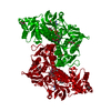
| ||||||||
| Unit cell |
|
-Components
-Protein , 1 types, 1 molecules A
| #1: Protein | Mass: 65061.145 Da / Num. of mol.: 1 Source method: isolated from a genetically manipulated source Source: (gene. exp.) SCHISTOSOMA MANSONI (invertebrata) / Plasmid: PGEX-4T1 / Production host:  ESCHERICHIA COLI (E. coli) / Strain (production host): BL21(DE3) / References: UniProt: Q962Y6, EC: 1.6.4.5 ESCHERICHIA COLI (E. coli) / Strain (production host): BL21(DE3) / References: UniProt: Q962Y6, EC: 1.6.4.5 |
|---|
-Non-polymers , 5 types, 263 molecules 


| #2: Chemical | ChemComp-FAD / Flavin adenine dinucleotide Mass: 785.550 Da / Num. of mol.: 1 / Source method: obtained synthetically / Formula: C27H33N9O15P2 / Comment: FAD*YM Mass: 785.550 Da / Num. of mol.: 1 / Source method: obtained synthetically / Formula: C27H33N9O15P2 / Comment: FAD*YM | ||||||
|---|---|---|---|---|---|---|---|
| #3: Chemical | ChemComp-PEG / Diethylene glycol Mass: 106.120 Da / Num. of mol.: 4 / Source method: obtained synthetically / Formula: C4H10O3 Mass: 106.120 Da / Num. of mol.: 4 / Source method: obtained synthetically / Formula: C4H10O3#4: Chemical | Glycerol Mass: 92.094 Da / Num. of mol.: 2 / Source method: obtained synthetically / Formula: C3H8O3 Mass: 92.094 Da / Num. of mol.: 2 / Source method: obtained synthetically / Formula: C3H8O3#5: Chemical | ChemComp-PG4 / | Polyethylene glycol Mass: 194.226 Da / Num. of mol.: 1 / Source method: obtained synthetically / Formula: C8H18O5 / Comment: precipitant*YM Mass: 194.226 Da / Num. of mol.: 1 / Source method: obtained synthetically / Formula: C8H18O5 / Comment: precipitant*YM#6: Water | ChemComp-HOH / | WaterMass: 18.015 Da / Num. of mol.: 255 / Source method: isolated from a natural source / Formula: H2O |
-Details
| Sequence details | RESIDUE 597 IS GIVEN AS X IN UNIPROT, IT IS CYS IN THIS CONSTRUCT |
|---|
-Experimental details
-Experiment
| Experiment | Method: X-RAY DIFFRACTION |
|---|
- Sample preparation
| Crystal | Density Matthews: 3.062555 Å3/Da / Density % sol: 63 % |
|---|---|
| Crystal grow | Method: vapor diffusion, sitting drop / pH: 7.4 Details: VAPOR DIFFUSION, SITTING DROPS, CRYSTALS GROWN IN HEPES 0.1 M PH 7.4, PEG 3350 20%, KI 0.2M, 2-MERCAPTOETHANOL 5MM SOAKED WITH CUSO4 0.001 MM. |
-Data collection
| Diffraction | Mean temperature: 100 K |
|---|---|
| Diffraction source | Source: SYNCHROTRON / Site: BESSY  / Type: BESSY / Wavelength: 0.918 / Type: BESSY / Wavelength: 0.918 |
| Radiation | Protocol: SINGLE WAVELENGTH / Monochromatic (M) / Laue (L): M / Scattering type: x-ray |
| Radiation wavelength | Wavelength: 0.918 Å / Relative weight: 1 |
| Reflection | Resolution: 1.9→40 Å / Num. obs: 52728 / % possible obs: 90 % / Observed criterion σ(I): 2 / Redundancy: 3 % / Biso Wilson estimate: 19 Å2 / Rmerge(I) obs: 0.07 / Net I/σ(I): 12.2 |
| Reflection shell | Resolution: 1.9→2 Å / Redundancy: 2.8 % / Rmerge(I) obs: 0.32 / Mean I/σ(I) obs: 3.2 / % possible all: 92.6 |
- Processing
| Software |
| ||||||||||||||||||||||||||||||||||||||||||||||||||||||||||||||||||||||||||||||||||||||||||||||||||||||||||||||||||||||||||||||||||||||||||||||||||||||||||||||||||||||||||||||||||||||
|---|---|---|---|---|---|---|---|---|---|---|---|---|---|---|---|---|---|---|---|---|---|---|---|---|---|---|---|---|---|---|---|---|---|---|---|---|---|---|---|---|---|---|---|---|---|---|---|---|---|---|---|---|---|---|---|---|---|---|---|---|---|---|---|---|---|---|---|---|---|---|---|---|---|---|---|---|---|---|---|---|---|---|---|---|---|---|---|---|---|---|---|---|---|---|---|---|---|---|---|---|---|---|---|---|---|---|---|---|---|---|---|---|---|---|---|---|---|---|---|---|---|---|---|---|---|---|---|---|---|---|---|---|---|---|---|---|---|---|---|---|---|---|---|---|---|---|---|---|---|---|---|---|---|---|---|---|---|---|---|---|---|---|---|---|---|---|---|---|---|---|---|---|---|---|---|---|---|---|---|---|---|---|---|
| Refinement | Method to determine structure: MOLECULAR REPLACEMENT Starting model: PDB ENTRY 2V6O Resolution: 1.9→40 Å / Cor.coef. Fo:Fc: 0.943 / Cor.coef. Fo:Fc free: 0.93 / SU B: 2.427 / SU ML: 0.073 / Cross valid method: THROUGHOUT / ESU R: 0.145 / ESU R Free: 0.128 / Stereochemistry target values: MAXIMUM LIKELIHOOD Details: HYDROGENS HAVE BEEN ADDED IN THE RIDING POSITIONS. RESIDUES 1-5 OF CHAIN A ARE NOT VISIBLE BY THE ELECTRON DENSITY MAPS
| ||||||||||||||||||||||||||||||||||||||||||||||||||||||||||||||||||||||||||||||||||||||||||||||||||||||||||||||||||||||||||||||||||||||||||||||||||||||||||||||||||||||||||||||||||||||
| Solvent computation | Ion probe radii: 0.8 Å / Shrinkage radii: 0.8 Å / VDW probe radii: 1.4 Å / Solvent model: MASK | ||||||||||||||||||||||||||||||||||||||||||||||||||||||||||||||||||||||||||||||||||||||||||||||||||||||||||||||||||||||||||||||||||||||||||||||||||||||||||||||||||||||||||||||||||||||
| Displacement parameters | Biso mean: 18.6 Å2
| ||||||||||||||||||||||||||||||||||||||||||||||||||||||||||||||||||||||||||||||||||||||||||||||||||||||||||||||||||||||||||||||||||||||||||||||||||||||||||||||||||||||||||||||||||||||
| Refinement step | Cycle: LAST / Resolution: 1.9→40 Å
| ||||||||||||||||||||||||||||||||||||||||||||||||||||||||||||||||||||||||||||||||||||||||||||||||||||||||||||||||||||||||||||||||||||||||||||||||||||||||||||||||||||||||||||||||||||||
| Refine LS restraints |
|