 Movie
Movie Controller

[English] 日本語
 Yorodumi
- PDB-2wz1: STRUCTURE OF THE CATALYTIC DOMAIN OF HUMAN SOLUBLE GUANYLATE CYCL... -
+ Open data

- Basic information
| Entry | Database: PDB / ID: 2wz1 | ||||||
|---|---|---|---|---|---|---|---|
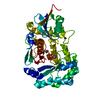
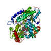
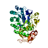
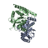
| Title | STRUCTURE OF THE CATALYTIC DOMAIN OF HUMAN SOLUBLE GUANYLATE CYCLASE 1 BETA 3. | ||||||
 Components Components | GUANYLATE CYCLASE SOLUBLE SUBUNIT BETA-1 | ||||||
 Keywords Keywords |  LYASE / GUCY1 / METAL-BINDING / CGMP BIOSYNTHESIS / NUCLEOTIDE-BINDING / CYCLASE / GUCY1B3 / GTP-BINDING LYASE / GUCY1 / METAL-BINDING / CGMP BIOSYNTHESIS / NUCLEOTIDE-BINDING / CYCLASE / GUCY1B3 / GTP-BINDING | ||||||
| Function / homology |  Function and homology informationcytidylate cyclase activity / guanylate cyclase complex, soluble / trans-synaptic signaling by nitric oxide, modulating synaptic transmission / guanylate cyclase / cGMP biosynthetic process / guanylate cyclase activity / response to oxygen levels / presynaptic active zone cytoplasmic component / Nitric oxide stimulates guanylate cyclase / adenylate cyclase activity ...cytidylate cyclase activity / guanylate cyclase complex, soluble / trans-synaptic signaling by nitric oxide, modulating synaptic transmission / guanylate cyclase / cGMP biosynthetic process / guanylate cyclase activity / response to oxygen levels / presynaptic active zone cytoplasmic component / Nitric oxide stimulates guanylate cyclase / adenylate cyclase activity / blood circulation / nitric oxide-cGMP-mediated signaling / cGMP-mediated signaling / Smooth Muscle Contraction / cellular response to nitric oxide / nitric oxide mediated signal transduction / Hsp90 protein binding / signaling receptor activity / glutamatergic synapse / heme binding / protein-containing complex binding / GTP binding / metal ion binding / cytosol Function and homology informationcytidylate cyclase activity / guanylate cyclase complex, soluble / trans-synaptic signaling by nitric oxide, modulating synaptic transmission / guanylate cyclase / cGMP biosynthetic process / guanylate cyclase activity / response to oxygen levels / presynaptic active zone cytoplasmic component / Nitric oxide stimulates guanylate cyclase / adenylate cyclase activity ...cytidylate cyclase activity / guanylate cyclase complex, soluble / trans-synaptic signaling by nitric oxide, modulating synaptic transmission / guanylate cyclase / cGMP biosynthetic process / guanylate cyclase activity / response to oxygen levels / presynaptic active zone cytoplasmic component / Nitric oxide stimulates guanylate cyclase / adenylate cyclase activity / blood circulation / nitric oxide-cGMP-mediated signaling / cGMP-mediated signaling / Smooth Muscle Contraction / cellular response to nitric oxide / nitric oxide mediated signal transduction / Hsp90 protein binding / signaling receptor activity / glutamatergic synapse / heme binding / protein-containing complex binding / GTP binding / metal ion binding / cytosolSimilarity search - Function | ||||||
| Biological species |  HOMO SAPIENS (human) HOMO SAPIENS (human) | ||||||
| Method | X-RAY DIFFRACTION / SYNCHROTRON / MOLECULAR REPLACEMENT / Resolution: 1.63 Å | ||||||
 Authors Authors | Allerston, C.K. / Cooper, C.D.O. / Muniz, J. / Pike, A.C.W. / von Delft, F. / Arrowsmith, C.H. / Weigelt, J. / Edwards, A. / Bountra, C. / Gileadi, O. | ||||||
 Citation Citation | Journal: Plos One / Year: 2013 Title: Crystal Structures of the Catalytic Domain of Human Soluble Guanylate Cyclase. Authors: Allerston, C.K. / von Delft, F. / Gileadi, O. | ||||||
| History |
|
- Structure visualization
| Structure viewer | Molecule: MolmilJmol/JSmol |
|---|
- Downloads & links
-Download
| PDBx/mmCIF format | 2wz1.cif.gz | 98.6 KB | Display | PDBx/mmCIF format |
|---|---|---|---|---|
| PDB format | pdb2wz1.ent.gz | 74.7 KB | Display | PDB format |
| PDBx/mmJSON format | 2wz1.json.gz | Tree view | PDBx/mmJSON format | |
| Others |  Other downloads Other downloads |
-Validation report
| Arichive directory | https://data.pdbj.org/pub/pdb/validation_reports/wz/2wz1ftp://data.pdbj.org/pub/pdb/validation_reports/wz/2wz1 | HTTPS FTP |
|---|
-Related structure data
| Related structure data |  3uvjC  3et6S S: Starting model for refinement C: citing same article ( |
|---|---|
| Similar structure data |


-Links
 PDBj
PDBj


- Assembly
| Deposited unit | 
| ||||||||
|---|---|---|---|---|---|---|---|---|---|
| 1 |
| ||||||||
| Unit cell |
| ||||||||
| Noncrystallographic symmetry (NCS) | NCS oper: (Code: given Matrix: (-0.94655, -0.08878, 0.3101), Vector : |
-Components
| #1: Protein | Mass: 24657.020 Da / Num. of mol.: 2 / Fragment: CATALYTIC DOMAIN, RESIDUES 994-1205 Source method: isolated from a genetically manipulated source Source: (gene. exp.) HOMO SAPIENS (human) / Plasmid: PNIC-CTHF / Production host:  ESCHERICHIA COLI (E. coli) / Strain (production host): BL21(DE3) / Variant (production host): R3 PRARE2 / References: UniProt: Q02153, guanylate cyclase ESCHERICHIA COLI (E. coli) / Strain (production host): BL21(DE3) / Variant (production host): R3 PRARE2 / References: UniProt: Q02153, guanylate cyclase#2: Chemical | Ethylene glycol  Mass: 62.068 Da / Num. of mol.: 3 / Source method: obtained synthetically / Formula: C2H6O2 Mass: 62.068 Da / Num. of mol.: 3 / Source method: obtained synthetically / Formula: C2H6O2#3: Water | ChemComp-HOH / | Water Mass: 18.015 Da / Num. of mol.: 289 / Source method: isolated from a natural source / Formula: H2O Mass: 18.015 Da / Num. of mol.: 289 / Source method: isolated from a natural source / Formula: H2O |
|---|
-Experimental details
-Experiment
| Experiment | Method: X-RAY DIFFRACTION / Number of used crystals: 1 |
|---|
- Sample preparation
| Crystal | Density Matthews: 2.1 Å3/Da / Density % sol: 41.45 % / Description: NONE |
|---|---|
| Crystal grow | Method: vapor diffusion, sitting drop / pH: 7.5 Details: PROTEIN WAS CONCENTRATED TO 12 MG/ML IN PURIFICATION BUFFER (10MM HEPES, 500MM NACL, 5% GLYCEROL, 10% GALACTOSE, 0.5MM TCEP)AND SET UP IN SITTING DROP IN A 1:2 RATIO WITH HAMPTON INDEX ...Details: PROTEIN WAS CONCENTRATED TO 12 MG/ML IN PURIFICATION BUFFER (10MM HEPES, 500MM NACL, 5% GLYCEROL, 10% GALACTOSE, 0.5MM TCEP)AND SET UP IN SITTING DROP IN A 1:2 RATIO WITH HAMPTON INDEX SCREEN - B8 (1.4M TRI-SODIUM CITRATE DIHYDRATE, 0.1M HEPES, PH 7.5). |
-Data collection
| Diffraction | Mean temperature: 287 K |
|---|---|
| Diffraction source | Source: SYNCHROTRON / Site: Diamond  / Beamline: I03 / Wavelength: 0.9795 / Beamline: I03 / Wavelength: 0.9795 |
| Detector | Type: ADSC CCD / Detector: CCD / Date: Oct 14, 2009 |
| Radiation | Protocol: SINGLE WAVELENGTH / Monochromatic (M) / Laue (L): M / Scattering type: x-ray |
| Radiation wavelength | Wavelength: 0.9795 Å / Relative weight: 1 |
| Reflection | Resolution: 1.63→37 Å / Num. obs: 49220 / % possible obs: 99.3 % / Observed criterion σ(I): 1.3 / Redundancy: 3.4 % / Biso Wilson estimate: 19.6 Å2 / Rmerge(I) obs: 0.06 / Net I/σ(I): 9.9 |
| Reflection shell | Resolution: 1.63→1.67 Å / Redundancy: 3.5 % / Rmerge(I) obs: 0.56 / Mean I/σ(I) obs: 2.1 / % possible all: 99.8 |
- Processing
| Software |
| ||||||||||||||||||||||||||||||||||||||||||||||||||||||||||||||||||||||||||||||||||||||||||||||||||||||||||||||||||||||||||||||||||||||||||||||||||||||||||||||||||||||||||||||||||||||
|---|---|---|---|---|---|---|---|---|---|---|---|---|---|---|---|---|---|---|---|---|---|---|---|---|---|---|---|---|---|---|---|---|---|---|---|---|---|---|---|---|---|---|---|---|---|---|---|---|---|---|---|---|---|---|---|---|---|---|---|---|---|---|---|---|---|---|---|---|---|---|---|---|---|---|---|---|---|---|---|---|---|---|---|---|---|---|---|---|---|---|---|---|---|---|---|---|---|---|---|---|---|---|---|---|---|---|---|---|---|---|---|---|---|---|---|---|---|---|---|---|---|---|---|---|---|---|---|---|---|---|---|---|---|---|---|---|---|---|---|---|---|---|---|---|---|---|---|---|---|---|---|---|---|---|---|---|---|---|---|---|---|---|---|---|---|---|---|---|---|---|---|---|---|---|---|---|---|---|---|---|---|---|---|
| Refinement | Method to determine structure: MOLECULAR REPLACEMENT Starting model: PDB ENTRY 3ET6 Resolution: 1.63→37 Å / Cor.coef. Fo:Fc: 0.961 / Cor.coef. Fo:Fc free: 0.948 / SU B: 4.37 / SU ML: 0.067 / TLS residual ADP flag: LIKELY RESIDUAL / Cross valid method: THROUGHOUT / ESU R: 0.095 / ESU R Free: 0.096 / Stereochemistry target values: MAXIMUM LIKELIHOOD Details: HYDROGENS HAVE BEEN ADDED IN THE RIDING POSITIONS. ATOM RECORD CONTAINS RESIDUAL B FACTORS ONLY.
| ||||||||||||||||||||||||||||||||||||||||||||||||||||||||||||||||||||||||||||||||||||||||||||||||||||||||||||||||||||||||||||||||||||||||||||||||||||||||||||||||||||||||||||||||||||||
| Solvent computation | Ion probe radii: 0.8 Å / Shrinkage radii: 0.8 Å / VDW probe radii: 1.4 Å / Solvent model: MASK | ||||||||||||||||||||||||||||||||||||||||||||||||||||||||||||||||||||||||||||||||||||||||||||||||||||||||||||||||||||||||||||||||||||||||||||||||||||||||||||||||||||||||||||||||||||||
| Displacement parameters | Biso mean: 12.388 Å2
| ||||||||||||||||||||||||||||||||||||||||||||||||||||||||||||||||||||||||||||||||||||||||||||||||||||||||||||||||||||||||||||||||||||||||||||||||||||||||||||||||||||||||||||||||||||||
| Refinement step | Cycle: LAST / Resolution: 1.63→37 Å
| ||||||||||||||||||||||||||||||||||||||||||||||||||||||||||||||||||||||||||||||||||||||||||||||||||||||||||||||||||||||||||||||||||||||||||||||||||||||||||||||||||||||||||||||||||||||
| Refine LS restraints |
|