 Movie
Movie Controller

[English] 日本語
 Yorodumi
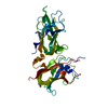
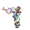
- PDB-2wuh: Crystal structure of the DDR2 discoidin domain bound to a triple-... -
+ Open data

- Basic information
| Entry | Database: PDB / ID: 2wuh | ||||||
|---|---|---|---|---|---|---|---|
| Title | Crystal structure of the DDR2 discoidin domain bound to a triple- helical collagen peptide | ||||||
 Components Components |
| ||||||
 Keywords Keywords | RECEPTOR/PEPTIDE / RECEPTOR-PEPTIDE COMPLEX /  TRANSFERASE / NUCLEOTIDE-BINDING / TYROSINE-PROTEIN KINASE TRANSFERASE / NUCLEOTIDE-BINDING / TYROSINE-PROTEIN KINASE | ||||||
| Function / homology |  Function and homology information Function and homology informationpositive regulation of hepatic stellate cell proliferation / biomineral tissue development / protein tyrosine kinase collagen receptor activity / positive regulation of extracellular matrix disassembly / chondrocyte proliferation / positive regulation of hepatic stellate cell activation / endochondral bone growth / regulation of extracellular matrix disassembly / negative regulation of hydrogen peroxide-mediated programmed cell death / collagen-activated tyrosine kinase receptor signaling pathway ...positive regulation of hepatic stellate cell proliferation / biomineral tissue development / protein tyrosine kinase collagen receptor activity / positive regulation of extracellular matrix disassembly / chondrocyte proliferation / positive regulation of hepatic stellate cell activation / endochondral bone growth / regulation of extracellular matrix disassembly / negative regulation of hydrogen peroxide-mediated programmed cell death / collagen-activated tyrosine kinase receptor signaling pathway / regulation of bone mineralization / positive regulation of vascular associated smooth muscle cell migration / positive regulation of fibroblast migration / collagen fibril organization / cellular response to angiotensin / regulation of tissue remodeling / positive regulation of wound healing / Non-integrin membrane-ECM interactions / positive regulation of collagen biosynthetic process / positive regulation of protein kinase activity / positive regulation of G1/S transition of mitotic cell cycle / positive regulation of osteoblast differentiation / positive regulation of vascular associated smooth muscle cell proliferation / cellular response to transforming growth factor beta stimulus / collagen binding / response to muscle stretch / transmembrane receptor protein tyrosine kinase activity / ossification / receptor protein-tyrosine kinase / positive regulation of DNA-binding transcription factor activity / positive regulation of neuron projection development / peptidyl-tyrosine phosphorylation / positive regulation of fibroblast proliferation / actin cytoskeleton / cellular response to hypoxia / protein autophosphorylation / receptor complex / cell adhesion / apical plasma membrane / focal adhesion / negative regulation of apoptotic process / signal transduction / ATP binding / plasma membraneSimilarity search - Function | ||||||
| Biological species |  HOMO SAPIENS (human) HOMO SAPIENS (human) | ||||||
| Method | X-RAY DIFFRACTION / SYNCHROTRON / MOLECULAR REPLACEMENT / Resolution: 1.6 Å | ||||||
 Authors Authors | Carafoli, F. / Bihan, D. / Stathopoulos, S. / Konitsiotis, A.D. / Kvansakul, M. / Farndale, R.W. / Leitinger, B. / Hohenester, E. | ||||||
 Citation Citation | Journal: Structure / Year: 2009 Title: Crystallographic Insight Into Collagen Recognition by Discoidin Domain Receptor 2 Authors: Carafoli, F. / Bihan, D. / Stathopoulos, S. / Konitsiotis, A.D. / Kvansakul, M. / Farndale, R.W. / Leitinger, B. / Hohenester, E. #1: Journal: Embo J. / Year: 2007Title: Structural Basis of the Collagen-Binding Mode of Discoidin Domain Receptor 2. Authors: Ichikawa, O. / Osawa, M. / Nishida, N. / Goshima, N. / Nomura, N. / Shimada, I. #2: Journal: J.Biol.Chem. / Year: 2008 Title: Characterization of High Affinity Binding Motifs for the Discoidin Domain Receptor Ddr2 in Collagen. Authors: Konitsiotis, A.D. / Raynal, N. / Bihan, D. / Hohenester, E. / Farndale, R.W. / Leitinger, B. | ||||||
| History |
| ||||||
| Remark 700 | SHEET THE SHEET STRUCTURE OF THIS MOLECULE IS BIFURCATED. IN ORDER TO REPRESENT THIS FEATURE IN ... SHEET THE SHEET STRUCTURE OF THIS MOLECULE IS BIFURCATED. IN ORDER TO REPRESENT THIS FEATURE IN THE SHEET RECORDS BELOW, TWO SHEETS ARE DEFINED. |
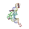
- Structure visualization
| Structure viewer | Molecule: MolmilJmol/JSmol |
|---|
- Downloads & links
-Download
| PDBx/mmCIF format | 2wuh.cif.gz | 65 KB | Display | PDBx/mmCIF format |
|---|---|---|---|---|
| PDB format | pdb2wuh.ent.gz | 48.1 KB | Display | PDB format |
| PDBx/mmJSON format | 2wuh.json.gz | Tree view | PDBx/mmJSON format | |
| Others |  Other downloads Other downloads |
-Validation report
| Arichive directory | https://data.pdbj.org/pub/pdb/validation_reports/wu/2wuhftp://data.pdbj.org/pub/pdb/validation_reports/wu/2wuh | HTTPS FTP |
|---|
-Related structure data


-Links
 PDBj
PDBj


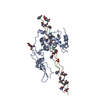
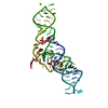
- Assembly
| Deposited unit | 
| ||||||||
|---|---|---|---|---|---|---|---|---|---|
| 1 |
| ||||||||
| Unit cell |
|
-Components
| #1: Protein | Mass: 20108.637 Da / Num. of mol.: 1 / Fragment: DISCOIDIN DOMAIN, RESIDUES 26-190 Source method: isolated from a genetically manipulated source Source: (gene. exp.) HOMO SAPIENS (human) / Cell line (production host): 293-EBNA / Production host: HOMO SAPIENS (human) / References: UniProt: Q16832 | ||||
|---|---|---|---|---|---|
| #2: Protein/peptide | Gelatin Mass: 2633.847 Da / Num. of mol.: 3 / Source method: obtained synthetically / Details: ACETYLATED N-TERMINUS / Source: (synth.) HOMO SAPIENS (human)#3: Water | ChemComp-HOH / | Water Mass: 18.015 Da / Num. of mol.: 176 / Source method: isolated from a natural source / Formula: H2O Mass: 18.015 Da / Num. of mol.: 176 / Source method: isolated from a natural source / Formula: H2ONonpolymer details | ACETYL GROUP (ACE): ACETYLATED | |
-Experimental details
-Experiment
| Experiment | Method: X-RAY DIFFRACTION / Number of used crystals: 1 |
|---|
- Sample preparation
| Crystal | Density Matthews: 2.1 Å3/Da / Density % sol: 40.85 % / Description: NONE |
|---|---|
| Crystal grow | pH: 7 / Details: pH 7.0 |
-Data collection
| Diffraction | Mean temperature: 100 K |
|---|---|
| Diffraction source | Source: SYNCHROTRON / Site: Diamond  / Beamline: I02 / Wavelength: 0.98 / Beamline: I02 / Wavelength: 0.98 |
| Detector | Detector: CCD / Date: Dec 16, 2008 |
| Radiation | Protocol: SINGLE WAVELENGTH / Monochromatic (M) / Laue (L): M / Scattering type: x-ray |
| Radiation wavelength | Wavelength: 0.98 Å / Relative weight: 1 |
| Reflection | Resolution: 1.6→20 Å / Num. obs: 27359 / % possible obs: 90.2 % / Observed criterion σ(I): 0 / Redundancy: 3.8 % / Rmerge(I) obs: 0.05 / Net I/σ(I): 16.8 |
| Reflection shell | Resolution: 1.6→1.69 Å / Redundancy: 3.8 % / Rmerge(I) obs: 0.28 / Mean I/σ(I) obs: 4.3 / % possible all: 67.4 |
- Processing
| Software |
| ||||||||||||||||||||||||||||||||||||||||||||||||||||||||||||||||||||||||||||||||
|---|---|---|---|---|---|---|---|---|---|---|---|---|---|---|---|---|---|---|---|---|---|---|---|---|---|---|---|---|---|---|---|---|---|---|---|---|---|---|---|---|---|---|---|---|---|---|---|---|---|---|---|---|---|---|---|---|---|---|---|---|---|---|---|---|---|---|---|---|---|---|---|---|---|---|---|---|---|---|---|---|---|
| Refinement | Method to determine structure: MOLECULAR REPLACEMENT Starting model: PDB ENTRIES 2QQJ AND 2V53 Resolution: 1.6→20 Å / Data cutoff high absF: 10000 / Data cutoff low absF: 0 / Isotropic thermal model: RESTRAINED / Cross valid method: THROUGHOUT / σ(F): 0 / Stereochemistry target values: MAXIMUM LIKELIHOOD
| ||||||||||||||||||||||||||||||||||||||||||||||||||||||||||||||||||||||||||||||||
| Solvent computation | Solvent model: FLAT MODEL / Bsol: 57.2491 Å2 / ksol: 0.45 e/Å3 | ||||||||||||||||||||||||||||||||||||||||||||||||||||||||||||||||||||||||||||||||
| Displacement parameters |
| ||||||||||||||||||||||||||||||||||||||||||||||||||||||||||||||||||||||||||||||||
| Refinement step | Cycle: LAST / Resolution: 1.6→20 Å
| ||||||||||||||||||||||||||||||||||||||||||||||||||||||||||||||||||||||||||||||||
| Refine LS restraints |
| ||||||||||||||||||||||||||||||||||||||||||||||||||||||||||||||||||||||||||||||||
| LS refinement shell | Resolution: 1.6→1.61 Å / Total num. of bins used: 50 /
| ||||||||||||||||||||||||||||||||||||||||||||||||||||||||||||||||||||||||||||||||
| Xplor file |
|
