 Movie
Movie Controller

[English] 日本語
 Yorodumi
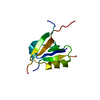
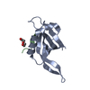
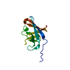
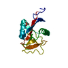
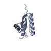
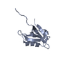
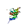
- PDB-2wt8: Structure of the N-terminal BRCT domain of human microcephalin (Mcph1) -
+ Open data

- Basic information
| Entry | Database: PDB / ID: 2wt8 | ||||||
|---|---|---|---|---|---|---|---|
| Title | Structure of the N-terminal BRCT domain of human microcephalin (Mcph1) | ||||||
 Components Components | MICROCEPHALIN | ||||||
 Keywords Keywords | CELL CYCLE / CHROMOSOME CONDENSATION / DWARFISM / POLYMORPHISM / MICROCEPHALY / PHOSPHOPROTEIN / MENTAL RETARDATION / PRIMARY MICROCEPHALY | ||||||
| Function / homology |  Function and homology information Function and homology informationregulation of chromosome condensation / neuronal stem cell population maintenance / regulation of centrosome cycle / protein localization to centrosome / establishment of mitotic spindle orientation / Condensation of Prophase Chromosomes / bone development / cerebral cortex development / mitotic cell cycle / regulation of inflammatory response ...regulation of chromosome condensation / neuronal stem cell population maintenance / regulation of centrosome cycle / protein localization to centrosome / establishment of mitotic spindle orientation / Condensation of Prophase Chromosomes / bone development / cerebral cortex development / mitotic cell cycle / regulation of inflammatory response / centrosome / negative regulation of transcription by RNA polymerase II / nucleoplasm / identical protein binding / cytoplasmSimilarity search - Function | ||||||
| Biological species |  HOMO SAPIENS (human) HOMO SAPIENS (human) | ||||||
| Method | X-RAY DIFFRACTION / SYNCHROTRON / SAD / Resolution: 1.6 Å | ||||||
 Authors Authors | Richards, M.W. / Roe, S.M. / Bayliss, R. | ||||||
 Citation Citation | Journal: J.Mol.Biol. / Year: 2010 Title: A Pocket on the Surface of the N-Terminal Brct Domain of Mcph1 is Required to Prevent Abnormal Chromosome Condensation. Authors: Richards, M.W. / Leung, J.W.C. / Roe, S.M. / Chen, J. / Bayliss, R. | ||||||
| History |
|
- Structure visualization
| Structure viewer | Molecule: MolmilJmol/JSmol |
|---|
- Downloads & links
-Download
| PDBx/mmCIF format | 2wt8.cif.gz | 162.5 KB | Display | PDBx/mmCIF format |
|---|---|---|---|---|
| PDB format | pdb2wt8.ent.gz | 137 KB | Display | PDB format |
| PDBx/mmJSON format | 2wt8.json.gz | Tree view | PDBx/mmJSON format | |
| Others |  Other downloads Other downloads |
-Validation report
| Arichive directory | https://data.pdbj.org/pub/pdb/validation_reports/wt/2wt8ftp://data.pdbj.org/pub/pdb/validation_reports/wt/2wt8 | HTTPS FTP |
|---|
-Related structure data
| Similar structure data |
|---|

-Links
 PDBj
PDBj


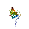
- Assembly
| Deposited unit | 
| ||||||||
|---|---|---|---|---|---|---|---|---|---|
| 1 | 
| ||||||||
| 2 | 
| ||||||||
| 3 | 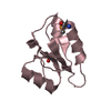
| ||||||||
| 4 | 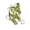
| ||||||||
| Unit cell |
|
-Components
| #1: Protein | Mass: 10818.010 Da / Num. of mol.: 4 / Fragment: N-TERMINAL BRCT DOMAIN, RESIDUES 1-95 Source method: isolated from a genetically manipulated source Source: (gene. exp.) HOMO SAPIENS (human) / Production host:  ESCHERICHIA COLI (E. coli) / Strain (production host): B834 / Variant (production host): CODONPLUS RPIL / References: UniProt: Q8NEM0 ESCHERICHIA COLI (E. coli) / Strain (production host): B834 / Variant (production host): CODONPLUS RPIL / References: UniProt: Q8NEM0#2: Chemical | Acetate  Mass: 59.044 Da / Num. of mol.: 2 / Source method: obtained synthetically / Formula: C2H3O2 Mass: 59.044 Da / Num. of mol.: 2 / Source method: obtained synthetically / Formula: C2H3O2#3: Chemical | ChemComp-CL / | Chloride  Mass: 35.453 Da / Num. of mol.: 1 / Source method: obtained synthetically / Formula: Cl Mass: 35.453 Da / Num. of mol.: 1 / Source method: obtained synthetically / Formula: Cl#4: Water | ChemComp-HOH / | Water Mass: 18.015 Da / Num. of mol.: 469 / Source method: isolated from a natural source / Formula: H2O Mass: 18.015 Da / Num. of mol.: 469 / Source method: isolated from a natural source / Formula: H2OSequence details | N-TERMINAL GA FROM VECTOR SEQUENCE | |
|---|
-Experimental details
-Experiment
| Experiment | Method: X-RAY DIFFRACTION |
|---|
- Sample preparation
| Crystal | Density Matthews: 2.5 Å3/Da / Density % sol: 50.77 % / Description: NONE |
|---|---|
| Crystal grow | Details: 100 MM MES, PH 6.0, 18% PEG 6000, 200 MM NH4CL |
-Data collection
| Diffraction | Mean temperature: 100 K |
|---|---|
| Diffraction source | Source: SYNCHROTRON / Site: Diamond  / Beamline: I03 / Wavelength: 0.92 / Beamline: I03 / Wavelength: 0.92 |
| Detector | Type: ADSC CCD / Detector: CCD |
| Radiation | Protocol: SINGLE WAVELENGTH / Monochromatic (M) / Laue (L): M / Scattering type: x-ray |
| Radiation wavelength | Wavelength: 0.92 Å / Relative weight: 1 |
| Reflection | Resolution: 1.6→45.27 Å / Num. obs: 56453 / % possible obs: 99.7 % / Observed criterion σ(I): 2 / Redundancy: 4.3 % / Biso Wilson estimate: 19.66 Å2 / Rmerge(I) obs: 0.05 / Net I/σ(I): 16.1 |
| Reflection shell | Resolution: 1.6→1.69 Å / Redundancy: 3.6 % / Rmerge(I) obs: 0.34 / Mean I/σ(I) obs: 3.2 / % possible all: 99.9 |
- Processing
| Software |
| |||||||||||||||||||||||||||||||||||||||||||||||||||||||||||||||||||||||||||||||||||||||||||||||||||||||||||||||||||||||||||||||||||||||||||||||||||
|---|---|---|---|---|---|---|---|---|---|---|---|---|---|---|---|---|---|---|---|---|---|---|---|---|---|---|---|---|---|---|---|---|---|---|---|---|---|---|---|---|---|---|---|---|---|---|---|---|---|---|---|---|---|---|---|---|---|---|---|---|---|---|---|---|---|---|---|---|---|---|---|---|---|---|---|---|---|---|---|---|---|---|---|---|---|---|---|---|---|---|---|---|---|---|---|---|---|---|---|---|---|---|---|---|---|---|---|---|---|---|---|---|---|---|---|---|---|---|---|---|---|---|---|---|---|---|---|---|---|---|---|---|---|---|---|---|---|---|---|---|---|---|---|---|---|---|---|---|
| Refinement | Method to determine structure: SAD Starting model: NONE Resolution: 1.6→45.248 Å / SU ML: 0.24 / σ(F): 0.01 / Phase error: 21.55 / Stereochemistry target values: ML
| |||||||||||||||||||||||||||||||||||||||||||||||||||||||||||||||||||||||||||||||||||||||||||||||||||||||||||||||||||||||||||||||||||||||||||||||||||
| Solvent computation | Shrinkage radii: 0.9 Å / VDW probe radii: 1.11 Å / Solvent model: FLAT BULK SOLVENT MODEL / Bsol: 49.822 Å2 / ksol: 0.357 e/Å3 | |||||||||||||||||||||||||||||||||||||||||||||||||||||||||||||||||||||||||||||||||||||||||||||||||||||||||||||||||||||||||||||||||||||||||||||||||||
| Displacement parameters | Biso mean: 28.02 Å2
| |||||||||||||||||||||||||||||||||||||||||||||||||||||||||||||||||||||||||||||||||||||||||||||||||||||||||||||||||||||||||||||||||||||||||||||||||||
| Refinement step | Cycle: LAST / Resolution: 1.6→45.248 Å
| |||||||||||||||||||||||||||||||||||||||||||||||||||||||||||||||||||||||||||||||||||||||||||||||||||||||||||||||||||||||||||||||||||||||||||||||||||
| Refine LS restraints |
| |||||||||||||||||||||||||||||||||||||||||||||||||||||||||||||||||||||||||||||||||||||||||||||||||||||||||||||||||||||||||||||||||||||||||||||||||||
| LS refinement shell |
| |||||||||||||||||||||||||||||||||||||||||||||||||||||||||||||||||||||||||||||||||||||||||||||||||||||||||||||||||||||||||||||||||||||||||||||||||||
| Refinement TLS params. | Method: refined / Refine-ID: X-RAY DIFFRACTION
| |||||||||||||||||||||||||||||||||||||||||||||||||||||||||||||||||||||||||||||||||||||||||||||||||||||||||||||||||||||||||||||||||||||||||||||||||||
| Refinement TLS group |
|
