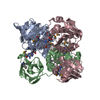
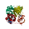
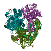
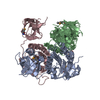
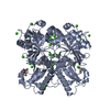
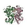
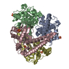
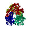
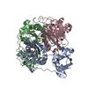
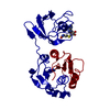
Entry Database : PDB / ID : 2wr8Title Structure of Pyrococcus horikoshii SAM hydroxide adenosyltransferase in complex with SAH PUTATIVE UNCHARACTERIZED PROTEIN PH0463 Keywords / / / / Function / homology Function Domain/homology Component
/ / / / / / / / / / / / / / / / / / / / / / / Biological species PYROCOCCUS HORIKOSHII (archaea)Method / / / Resolution : 1.77 Å Authors McMahon, S.A. / Deng, H. / O'Hagan, D. / Johnson, K.A. / Naismith, J.H. Journal : Chembiochem / Year : 2009Title : Mechanistic Insights Into Water Activation in Sam Hydroxide Adenosyltransferase (Duf-62).Authors : Deng, H. / Mcmahon, S.A. / Eustaquio, A.S. / Moore, B.S. / Naismith, J.H. / O'Hagan, D. History Deposition Aug 31, 2009 Deposition site / Processing site Revision 1.0 Sep 22, 2009 Provider / Type Revision 1.1 Jul 13, 2011 Group / Version format complianceRevision 1.2 Dec 20, 2023 Group Data collection / Database references ... Data collection / Database references / Derived calculations / Other / Refinement description Category chem_comp_atom / chem_comp_bond ... chem_comp_atom / chem_comp_bond / database_2 / pdbx_database_status / pdbx_initial_refinement_model / struct_site Item _database_2.pdbx_DOI / _database_2.pdbx_database_accession ... _database_2.pdbx_DOI / _database_2.pdbx_database_accession / _pdbx_database_status.status_code_sf / _struct_site.pdbx_auth_asym_id / _struct_site.pdbx_auth_comp_id / _struct_site.pdbx_auth_seq_id
Show all Show less Remark 700 SHEET THE SHEET STRUCTURE OF THIS MOLECULE IS BIFURCATED. IN ORDER TO REPRESENT THIS FEATURE IN ... SHEET THE SHEET STRUCTURE OF THIS MOLECULE IS BIFURCATED. IN ORDER TO REPRESENT THIS FEATURE IN THE SHEET RECORDS BELOW, TWO SHEETS ARE DEFINED.
 Movie
Movie Controller

 Yorodumi
 Open data
 Basic information
 Components
 Keywords
 TRANSFERASE / SAM / SAM HYDROXIDE ADENOSYLTRANSFERASE (DUF-62) / SN2 / WATER ACTIVATION
 Function and homology information

 Authors
 Citation
 Structure visualization
 Downloads & links
 Other downloads
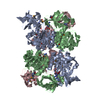

 PDBj
PDBj Assembly
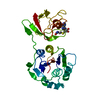


 Type: L-peptide linking / Mass: 384.411 Da / Num. of mol.: 1 / Source method: obtained synthetically / Formula: C14H20N6O5S
 Mass: 18.015 Da / Num. of mol.: 115 / Source method: isolated from a natural source / Formula: H2O
 Sample preparation
 / Beamline: ID14-4 / Wavelength: 0.98
  Processing