 Movie
Movie Controller

[English] 日本語
 Yorodumi
- PDB-2wcj: Structure of BMori GOBP2 (General Odorant Binding Protein 2) with... -
+ Open data

- Basic information
| Entry | Database: PDB / ID: 2wcj | ||||||
|---|---|---|---|---|---|---|---|
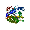
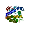
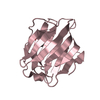
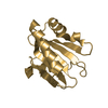
| Title | Structure of BMori GOBP2 (General Odorant Binding Protein 2) with (10E,12Z)-tetradecadien-1-ol | ||||||
 Components Components | GENERAL ODORANT-BINDING PROTEIN 1 | ||||||
 Keywords Keywords |  TRANSPORT PROTEIN / ODORANT BINDING PROTEIN / OLFACTION / TRANSPORT / DISULFIDE BOND / INSECT PHEREMONE / SENSORY TRANSDUCTION TRANSPORT PROTEIN / ODORANT BINDING PROTEIN / OLFACTION / TRANSPORT / DISULFIDE BOND / INSECT PHEREMONE / SENSORY TRANSDUCTION | ||||||
| Function / homology |  Function and homology information Function and homology information | ||||||
| Biological species |  BOMBYX MORI (domestic silkworm) BOMBYX MORI (domestic silkworm) | ||||||
| Method | X-RAY DIFFRACTION / SYNCHROTRON / MOLECULAR REPLACEMENT / Resolution: 1.4 Å | ||||||
 Authors Authors | Robertson, G. / Zhou, J.-J. / He, X. / Pickett, J.A. / Field, L.M. / Keep, N.H. | ||||||
 Citation Citation | Journal: J.Mol.Biol. / Year: 2009 Title: Characterisation of Bombyx Mori Odorant-Binding Proteins Reveals that a General Odorant-Binding Protein Discriminates between Sex Pheromone Components. Authors: Zhou, J.-J. / Robertson, G. / He, X. / Dufour, S. / Hooper, A.M. / Pickett, J.A. / Keep, N.H. / Field, L.M. | ||||||
| History |
|
- Structure visualization
| Structure viewer | Molecule: MolmilJmol/JSmol |
|---|
- Downloads & links
-Download
| PDBx/mmCIF format | 2wcj.cif.gz | 50.1 KB | Display | PDBx/mmCIF format |
|---|---|---|---|---|
| PDB format | pdb2wcj.ent.gz | 34.4 KB | Display | PDB format |
| PDBx/mmJSON format | 2wcj.json.gz | Tree view | PDBx/mmJSON format | |
| Others |  Other downloads Other downloads |
-Validation report
| Arichive directory | https://data.pdbj.org/pub/pdb/validation_reports/wc/2wcjftp://data.pdbj.org/pub/pdb/validation_reports/wc/2wcj | HTTPS FTP |
|---|
-Related structure data
| Related structure data |  2wc5SC  2wc6C  2wchC  2wckC  2wclC  2wcmC C: citing same article ( S: Starting model for refinement |
|---|---|
| Similar structure data |


-Links
 PDBj
PDBj- Assembly
| Deposited unit | 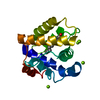
| ||||||||
|---|---|---|---|---|---|---|---|---|---|
| 1 |
| ||||||||
| Unit cell |
|
-Components
| #1: Protein | Mass: 16187.409 Da / Num. of mol.: 1 / Fragment: RESIDUES 20-160 Source method: isolated from a genetically manipulated source Source: (gene. exp.) BOMBYX MORI (domestic silkworm) / Organ: ANTENNA / Plasmid: PET17B / Production host:  ESCHERICHIA COLI (E. coli) / Strain (production host): BL21(DE3) / References: UniProt: P34170 ESCHERICHIA COLI (E. coli) / Strain (production host): BL21(DE3) / References: UniProt: P34170 | ||||||
|---|---|---|---|---|---|---|---|
| #2: Chemical | ChemComp-MG /   Mass: 24.305 Da / Num. of mol.: 6 / Source method: obtained synthetically / Formula: Mg Mass: 24.305 Da / Num. of mol.: 6 / Source method: obtained synthetically / Formula: Mg#3: Chemical | ChemComp-M21 / ( |   Mass: 210.356 Da / Num. of mol.: 1 / Source method: obtained synthetically / Formula: C14H26O Mass: 210.356 Da / Num. of mol.: 1 / Source method: obtained synthetically / Formula: C14H26O#4: Water | ChemComp-HOH / | Water Mass: 18.015 Da / Num. of mol.: 135 / Source method: isolated from a natural source / Formula: H2O Mass: 18.015 Da / Num. of mol.: 135 / Source method: isolated from a natural source / Formula: H2OSequence details | THREE POINT MUTATIONS DUE TO STRAIN DIFFERENCES. THE CDNA USED FOR THIS EXPERIMENT WAS OBTAINED ...THREE POINT MUTATIONS DUE TO STRAIN DIFFERENCE | |
-Experimental details
-Experiment
| Experiment | Method: X-RAY DIFFRACTION / Number of used crystals: 1 |
|---|
- Sample preparation
| Crystal | Density Matthews: 1.8 Å3/Da / Density % sol: 33 % / Description: NONE |
|---|---|
| Crystal grow | pH: 8.5 / Details: 30% PEG 4000, 200MM MGCL2, 100MM TRIS PH 8.5 |
-Data collection
| Diffraction | Mean temperature: 100 K |
|---|---|
| Diffraction source | Source: SYNCHROTRON / Site: Diamond  / Beamline: I04 / Wavelength: 0.9786 / Beamline: I04 / Wavelength: 0.9786 |
| Detector | Type: ADSC CCD / Detector: CCD / Date: May 14, 2008 |
| Radiation | Protocol: SINGLE WAVELENGTH / Monochromatic (M) / Laue (L): M / Scattering type: x-ray |
| Radiation wavelength | Wavelength: 0.9786 Å / Relative weight: 1 |
| Reflection | Resolution: 1.4→57.5 Å / Num. obs: 19390 / % possible obs: 94.7 % / Observed criterion σ(I): 0 / Redundancy: 6 % / Rmerge(I) obs: 0.07 / Net I/σ(I): 13.3 |
| Reflection shell | Resolution: 1.4→1.48 Å / Redundancy: 4.6 % / Rmerge(I) obs: 0.25 / Mean I/σ(I) obs: 4.8 / % possible all: 89.6 |
- Processing
| Software |
| ||||||||||||||||||||||||||||||||||||||||||||||||||||||||||||||||||||||||||||||||||||||||||||||||||||||||||||||||||||||||||||||||||||||||||||||||||||||||||||||||||||||||||||||||||||||
|---|---|---|---|---|---|---|---|---|---|---|---|---|---|---|---|---|---|---|---|---|---|---|---|---|---|---|---|---|---|---|---|---|---|---|---|---|---|---|---|---|---|---|---|---|---|---|---|---|---|---|---|---|---|---|---|---|---|---|---|---|---|---|---|---|---|---|---|---|---|---|---|---|---|---|---|---|---|---|---|---|---|---|---|---|---|---|---|---|---|---|---|---|---|---|---|---|---|---|---|---|---|---|---|---|---|---|---|---|---|---|---|---|---|---|---|---|---|---|---|---|---|---|---|---|---|---|---|---|---|---|---|---|---|---|---|---|---|---|---|---|---|---|---|---|---|---|---|---|---|---|---|---|---|---|---|---|---|---|---|---|---|---|---|---|---|---|---|---|---|---|---|---|---|---|---|---|---|---|---|---|---|---|---|
| Refinement | Method to determine structure: MOLECULAR REPLACEMENT Starting model: PDB ENTRY 2WC5 Resolution: 1.4→57.5 Å / Cor.coef. Fo:Fc: 0.957 / Cor.coef. Fo:Fc free: 0.904 / SU B: 1.188 / SU ML: 0.049 / Cross valid method: THROUGHOUT / ESU R: 0.077 / ESU R Free: 0.086 / Stereochemistry target values: MAXIMUM LIKELIHOOD / Details: HYDROGENS HAVE BEEN ADDED IN THE RIDING POSITIONS.
| ||||||||||||||||||||||||||||||||||||||||||||||||||||||||||||||||||||||||||||||||||||||||||||||||||||||||||||||||||||||||||||||||||||||||||||||||||||||||||||||||||||||||||||||||||||||
| Solvent computation | Ion probe radii: 0.8 Å / Shrinkage radii: 0.8 Å / VDW probe radii: 1.4 Å / Solvent model: MASK | ||||||||||||||||||||||||||||||||||||||||||||||||||||||||||||||||||||||||||||||||||||||||||||||||||||||||||||||||||||||||||||||||||||||||||||||||||||||||||||||||||||||||||||||||||||||
| Displacement parameters | Biso mean: 14.1 Å2
| ||||||||||||||||||||||||||||||||||||||||||||||||||||||||||||||||||||||||||||||||||||||||||||||||||||||||||||||||||||||||||||||||||||||||||||||||||||||||||||||||||||||||||||||||||||||
| Refinement step | Cycle: LAST / Resolution: 1.4→57.5 Å
| ||||||||||||||||||||||||||||||||||||||||||||||||||||||||||||||||||||||||||||||||||||||||||||||||||||||||||||||||||||||||||||||||||||||||||||||||||||||||||||||||||||||||||||||||||||||
| Refine LS restraints |
|