 Movie
Movie Controller


+ Open data

- Basic information
| Entry | Database: PDB / ID: 2wcb | ||||||
|---|---|---|---|---|---|---|---|
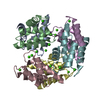
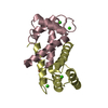
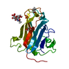
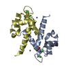
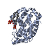
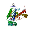
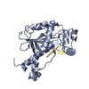
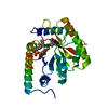
| Title | S100A12 complex with zinc in the absence of calcium | ||||||
 Components Components | PROTEIN S100-A12 | ||||||
 Keywords Keywords | METAL BINDING PROTEIN /  CALCIUM SIGNALLING / HOST-PARASITE RESPONSE CALCIUM SIGNALLING / HOST-PARASITE RESPONSE | ||||||
| Function / homology |  Function and homology information Function and homology informationmast cell activation / RAGE receptor binding / monocyte chemotaxis / TRAF6 mediated NF-kB activation / Advanced glycosylation endproduct receptor signaling / endothelial cell migration / defense response to fungus / xenobiotic metabolic process / neutrophil chemotaxis / positive regulation of MAP kinase activity ...mast cell activation / RAGE receptor binding / monocyte chemotaxis / TRAF6 mediated NF-kB activation / Advanced glycosylation endproduct receptor signaling / endothelial cell migration / defense response to fungus / xenobiotic metabolic process / neutrophil chemotaxis / positive regulation of MAP kinase activity / TAK1-dependent IKK and NF-kappa-B activation / positive regulation of inflammatory response / calcium-dependent protein binding / antimicrobial humoral immune response mediated by antimicrobial peptide / positive regulation of NF-kappaB transcription factor activity / secretory granule lumen / positive regulation of canonical NF-kappaB signal transduction / killing of cells of another organism / cytoskeleton / defense response to bacterium / inflammatory response / copper ion binding / innate immune response / calcium ion binding / Neutrophil degranulation / extracellular space / zinc ion binding / extracellular region / identical protein binding / nucleus / plasma membrane / cytosol / cytoplasmSimilarity search - Function | ||||||
| Biological species |  HOMO SAPIENS (human) HOMO SAPIENS (human) | ||||||
| Method | X-RAY DIFFRACTION / SYNCHROTRON / MOLECULAR REPLACEMENT / Resolution: 1.73 Å | ||||||
 Authors Authors | Moroz, O.V. / Blagova, E.V. / Wilkinson, A.J. / Wilson, K.S. / Bronstein, I.B. | ||||||
 Citation Citation | Journal: J.Mol.Biol. / Year: 2009 Title: The Crystal Structures of Human S100A12 in Apo Form and in Complex with Zinc: New Insights Into S100A12 Oligomerisation. Authors: Moroz, O.V. / Blagova, E.V. / Wilkinson, A.J. / Wilson, K.S. / Bronstein, I.B. #1: Journal: Acta Crystallogr.,Sect.D / Year: 2001Title: The Three-Dimensional Structure of Human S100A12. Authors: Moroz, O.V. / Antson, A.A. / Murshudov, G.N. / Maitland, N.J. / Dodson, G.G. / Wilson, K.S. / Skibshoj, I. / Lukanidin, E.M. / Bronstein, I.B. #2: Journal: Acta Crystallogr.,Sect.D / Year: 2003Title: Structure of the Human S100A12-Copper Complex: Implications for Host-Parasite Defence. Authors: Moroz, O.V. / Antson, A.A. / Grist, S.J. / Maitland, N.J. / Dodson, G.G. / Wilson, K.S. / Lukanidin, E. / Bronstein, I.B. #3: Journal: Acta Crystallogr.,Sect.D / Year: 2002Title: The Structure of S100A12 in a Hexameric Form and its Proposed Role in Receptor Signalling. Authors: Moroz, O.V. / Antson, A.A. / Dodson, E.J. / Burrell, H.J. / Grist, S.J. / Lloyd, R.M. / Maitland, N.J. / Dodson, G.G. / Wilson, K.S. / Lukanidin, E. / Bronstein, I.B. | ||||||
| History |
|
- Structure visualization
| Structure viewer | Molecule: MolmilJmol/JSmol |
|---|
- Downloads & links
-Download
| PDBx/mmCIF format | 2wcb.cif.gz | 56.4 KB | Display | PDBx/mmCIF format |
|---|---|---|---|---|
| PDB format | pdb2wcb.ent.gz | 41.1 KB | Display | PDB format |
| PDBx/mmJSON format | 2wcb.json.gz | Tree view | PDBx/mmJSON format | |
| Others |  Other downloads Other downloads |
-Validation report
| Arichive directory | https://data.pdbj.org/pub/pdb/validation_reports/wc/2wcbftp://data.pdbj.org/pub/pdb/validation_reports/wc/2wcb | HTTPS FTP |
|---|
-Related structure data
| Related structure data |  2wc8C 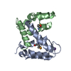 2wceC  2wcfC  1e8aS C: citing same article ( S: Starting model for refinement |
|---|---|
| Similar structure data |


-Links
 PDBj
PDBj


- Assembly
| Deposited unit | 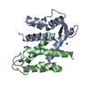
| ||||||||
|---|---|---|---|---|---|---|---|---|---|
| 1 |
| ||||||||
| Unit cell |
|
-Components
| #1: Protein | Mass: 10793.212 Da / Num. of mol.: 2 / Fragment: RESIDUES 2-92 Source method: isolated from a genetically manipulated source Source: (gene. exp.) HOMO SAPIENS (human) / Plasmid: PQE60 / Production host:  ESCHERICHIA COLI (E. coli) / References: UniProt: P80511 ESCHERICHIA COLI (E. coli) / References: UniProt: P80511#2: Chemical |   Mass: 65.409 Da / Num. of mol.: 2 / Source method: obtained synthetically / Formula: Zn Mass: 65.409 Da / Num. of mol.: 2 / Source method: obtained synthetically / Formula: Zn#3: Chemical |   Mass: 22.990 Da / Num. of mol.: 2 / Source method: obtained synthetically / Formula: Na Mass: 22.990 Da / Num. of mol.: 2 / Source method: obtained synthetically / Formula: Na#4: Water | ChemComp-HOH / | Water Mass: 18.015 Da / Num. of mol.: 166 / Source method: isolated from a natural source / Formula: H2O Mass: 18.015 Da / Num. of mol.: 166 / Source method: isolated from a natural source / Formula: H2OSequence details | EXTRA FOUR RESIDUES AT N TERMINUS MGGS | |
|---|
-Experimental details
-Experiment
| Experiment | Method: X-RAY DIFFRACTION / Number of used crystals: 1 |
|---|
- Sample preparation
| Crystal | Density Matthews: 3.21 Å3/Da / Density % sol: 61 % / Description: NONE |
|---|---|
| Crystal grow | pH: 7.5 Details: 34% PEG 3350, MME, 0.2M TRI AMMONIUM CITRATE PH 7.0 |
-Data collection
| Diffraction | Mean temperature: 120 K |
|---|---|
| Diffraction source | Source: SYNCHROTRON / Site: ESRF  / Beamline: BM14 / Wavelength: 1.25 / Beamline: BM14 / Wavelength: 1.25 |
| Detector | Type: MARMOSAIC 225 mm CCD / Detector: CCD |
| Radiation | Protocol: SINGLE WAVELENGTH / Monochromatic (M) / Laue (L): M / Scattering type: x-ray |
| Radiation wavelength | Wavelength: 1.25 Å / Relative weight: 1 |
| Reflection | Resolution: 1.73→77.8 Å / Num. obs: 28275 / % possible obs: 99.6 % / Observed criterion σ(I): 2 / Redundancy: 5.1 % / Rmerge(I) obs: 0.08 / Net I/σ(I): 17.1 |
| Reflection shell | Highest resolution: 1.73 Å / Redundancy: 3.7 % / Rmerge(I) obs: 0.72 / Mean I/σ(I) obs: 1.7 / % possible all: 99.6 |
- Processing
| Software |
| ||||||||||||||||||||||||||||||||||||||||||||||||||||||||||||||||||||||||||||||||||||||||||||||||||||||||||||||||||||||||||||||||||||||||||||||||||||||||||||||||||||||||||||||||||||||
|---|---|---|---|---|---|---|---|---|---|---|---|---|---|---|---|---|---|---|---|---|---|---|---|---|---|---|---|---|---|---|---|---|---|---|---|---|---|---|---|---|---|---|---|---|---|---|---|---|---|---|---|---|---|---|---|---|---|---|---|---|---|---|---|---|---|---|---|---|---|---|---|---|---|---|---|---|---|---|---|---|---|---|---|---|---|---|---|---|---|---|---|---|---|---|---|---|---|---|---|---|---|---|---|---|---|---|---|---|---|---|---|---|---|---|---|---|---|---|---|---|---|---|---|---|---|---|---|---|---|---|---|---|---|---|---|---|---|---|---|---|---|---|---|---|---|---|---|---|---|---|---|---|---|---|---|---|---|---|---|---|---|---|---|---|---|---|---|---|---|---|---|---|---|---|---|---|---|---|---|---|---|---|---|
| Refinement | Method to determine structure: MOLECULAR REPLACEMENT Starting model: PDB ENTRY 1E8A Resolution: 1.73→77.85 Å / Cor.coef. Fo:Fc: 0.953 / Cor.coef. Fo:Fc free: 0.94 / SU B: 8.668 / SU ML: 0.119 / TLS residual ADP flag: LIKELY RESIDUAL / Cross valid method: THROUGHOUT / ESU R: 0.116 / ESU R Free: 0.116 / Stereochemistry target values: MAXIMUM LIKELIHOOD Details: HYDROGENS HAVE BEEN ADDED IN THE RIDING POSITIONS. U VALUES RESIDUAL ONLY
| ||||||||||||||||||||||||||||||||||||||||||||||||||||||||||||||||||||||||||||||||||||||||||||||||||||||||||||||||||||||||||||||||||||||||||||||||||||||||||||||||||||||||||||||||||||||
| Solvent computation | Ion probe radii: 0.8 Å / Shrinkage radii: 0.8 Å / VDW probe radii: 1.2 Å / Solvent model: MASK | ||||||||||||||||||||||||||||||||||||||||||||||||||||||||||||||||||||||||||||||||||||||||||||||||||||||||||||||||||||||||||||||||||||||||||||||||||||||||||||||||||||||||||||||||||||||
| Displacement parameters | Biso mean: 11.11 Å2
| ||||||||||||||||||||||||||||||||||||||||||||||||||||||||||||||||||||||||||||||||||||||||||||||||||||||||||||||||||||||||||||||||||||||||||||||||||||||||||||||||||||||||||||||||||||||
| Refinement step | Cycle: LAST / Resolution: 1.73→77.85 Å
| ||||||||||||||||||||||||||||||||||||||||||||||||||||||||||||||||||||||||||||||||||||||||||||||||||||||||||||||||||||||||||||||||||||||||||||||||||||||||||||||||||||||||||||||||||||||
| Refine LS restraints |
|