 Movie
Movie Controller

[English] 日本語
 Yorodumi
- PDB-2wbt: The Structure of a Double C2H2 Zinc Finger Protein from a Hyperth... -
+ Open data

- Basic information
| Entry | Database: PDB / ID: 2wbt | ||||||
|---|---|---|---|---|---|---|---|
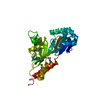
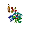
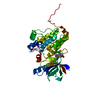
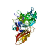
| Title | The Structure of a Double C2H2 Zinc Finger Protein from a Hyperthermophilic Archaeal Virus in the Absence of DNA | ||||||
 Components Components | B-129 | ||||||
 Keywords Keywords |  DNA BINDING PROTEIN / DNA-BINDING PROTEIN / ZINC FINGER / METAL-BINDING / DNA-BINDING PROTEIN SSV DNA BINDING PROTEIN / DNA-BINDING PROTEIN / ZINC FINGER / METAL-BINDING / DNA-BINDING PROTEIN SSV | ||||||
| Function / homology |  Function and homology information Function and homology information | ||||||
| Biological species |   SULFOLOBUS VIRUS 1 SULFOLOBUS VIRUS 1 | ||||||
| Method | X-RAY DIFFRACTION / SYNCHROTRON / MAD / Resolution: 2.7 Å | ||||||
 Authors Authors | Eilers, B.J. / Menon, S. / Windham, A.B. / Kraft, P. / Dlakic, M. / Young, M.J. / Lawrence, C.M. | ||||||
 Citation Citation | Journal: To be Published Title: The Structure of a Double C2H2 Zinc Finger Protein from a Hyperthermophilic Archaeal Virus in the Absence of DNA Authors: Eilers, B.J. / Menon, S. / Windham, A.B. / Kraft, P. / Dlakic, M. / Young, M.J. / Lawrence, C.M. | ||||||
| History |
|
- Structure visualization
| Structure viewer | Molecule: MolmilJmol/JSmol |
|---|
- Downloads & links
-Download
| PDBx/mmCIF format | 2wbt.cif.gz | 55.3 KB | Display | PDBx/mmCIF format |
|---|---|---|---|---|
| PDB format | pdb2wbt.ent.gz | 45.5 KB | Display | PDB format |
| PDBx/mmJSON format | 2wbt.json.gz | Tree view | PDBx/mmJSON format | |
| Others |  Other downloads Other downloads |
-Validation report
| Arichive directory | https://data.pdbj.org/pub/pdb/validation_reports/wb/2wbtftp://data.pdbj.org/pub/pdb/validation_reports/wb/2wbt | HTTPS FTP |
|---|
-Related structure data
| Similar structure data |
|---|


-Links
 PDBj
PDBj


- Assembly
| Deposited unit | 
| |||||||||||||||||||||||||||||||||||||||||||||||||||||||||||||||||||||||||||||||||||||||||||||||||||
|---|---|---|---|---|---|---|---|---|---|---|---|---|---|---|---|---|---|---|---|---|---|---|---|---|---|---|---|---|---|---|---|---|---|---|---|---|---|---|---|---|---|---|---|---|---|---|---|---|---|---|---|---|---|---|---|---|---|---|---|---|---|---|---|---|---|---|---|---|---|---|---|---|---|---|---|---|---|---|---|---|---|---|---|---|---|---|---|---|---|---|---|---|---|---|---|---|---|---|---|---|
| 1 |
| |||||||||||||||||||||||||||||||||||||||||||||||||||||||||||||||||||||||||||||||||||||||||||||||||||
| Unit cell |
| |||||||||||||||||||||||||||||||||||||||||||||||||||||||||||||||||||||||||||||||||||||||||||||||||||
| Noncrystallographic symmetry (NCS) | NCS domain:
NCS domain segments:
|
-Components
| #1: Protein | Mass: 14830.291 Da / Num. of mol.: 2 Source method: isolated from a genetically manipulated source Details: DISULFIDE BOND BETWEEN C 121 AND C 127 / Source: (gene. exp.) SULFOLOBUS VIRUS 1 / Plasmid: PEXP14-B129 / Production host:  ESCHERICHIA COLI (E. coli) / Strain (production host): BL21(DE3)RIL / References: UniProt: P20201 ESCHERICHIA COLI (E. coli) / Strain (production host): BL21(DE3)RIL / References: UniProt: P20201#2: Chemical | ChemComp-ZN /   Mass: 65.409 Da / Num. of mol.: 4 / Source method: obtained synthetically / Formula: Zn Mass: 65.409 Da / Num. of mol.: 4 / Source method: obtained synthetically / Formula: Zn#3: Water | ChemComp-HOH / | Water Mass: 18.015 Da / Num. of mol.: 17 / Source method: isolated from a natural source / Formula: H2O Mass: 18.015 Da / Num. of mol.: 17 / Source method: isolated from a natural source / Formula: H2O |
|---|
-Experimental details
-Experiment
| Experiment | Method: X-RAY DIFFRACTION / Number of used crystals: 1 |
|---|
- Sample preparation
| Crystal | Density Matthews: 2.7 Å3/Da / Density % sol: 0.55 % / Description: NONE |
|---|---|
| Crystal grow | Method: vapor diffusion, sitting drop / pH: 5.3 Details: SITTING DROP VAPOR DIFFUSION, 20 MG/ML OF B129 IN 20 MM BIS-TRIS, PH 6.5, 60 MM NACL MIXED WITH 0.1 M AMMONIUM CITRATE DIBASIC, 10-14 % PEG 4000 |
-Data collection
| Diffraction | Mean temperature: 110 K | ||||||||||||
|---|---|---|---|---|---|---|---|---|---|---|---|---|---|
| Diffraction source | Source: SYNCHROTRON / Site: SSRL  / Beamline: BL9-2 / Wavelength: 1.2833, 1.2830, 1.1696 / Beamline: BL9-2 / Wavelength: 1.2833, 1.2830, 1.1696 | ||||||||||||
| Detector | Type: MARRESEARCH / Detector: CCD / Date: Mar 18, 2006 Details: FLAT COLLIMATING MIRROR, DOUBLE CRYSTAL MONOCHROMATOR, TOROID FOCUSING MIRROR | ||||||||||||
| Radiation | Monochromator: DOUBLE CRYSTAL MONOCHROMATOR / Protocol: MAD / Monochromatic (M) / Laue (L): M / Scattering type: x-ray | ||||||||||||
| Radiation wavelength |
| ||||||||||||
| Reflection | Resolution: 2.7→50 Å / Num. obs: 8209 / % possible obs: 78.7 % / Observed criterion σ(I): 5 / Redundancy: 2.9 % / Rmerge(I) obs: 0.07 / Net I/σ(I): 17.2 | ||||||||||||
| Reflection shell | Resolution: 2.7→2.77 Å / Redundancy: 2.8 % / Rmerge(I) obs: 0.24 / Mean I/σ(I) obs: 4.4 / % possible all: 78.8 |
- Processing
| Software |
| ||||||||||||||||||||||||||||||||||||||||||||||||||||||||||||||||||||||||||||||||||||||||||||||||||||||||||||||||||||||||||||||||||||||||||||||||||||||||||||||||||||||||||||||||||||||
|---|---|---|---|---|---|---|---|---|---|---|---|---|---|---|---|---|---|---|---|---|---|---|---|---|---|---|---|---|---|---|---|---|---|---|---|---|---|---|---|---|---|---|---|---|---|---|---|---|---|---|---|---|---|---|---|---|---|---|---|---|---|---|---|---|---|---|---|---|---|---|---|---|---|---|---|---|---|---|---|---|---|---|---|---|---|---|---|---|---|---|---|---|---|---|---|---|---|---|---|---|---|---|---|---|---|---|---|---|---|---|---|---|---|---|---|---|---|---|---|---|---|---|---|---|---|---|---|---|---|---|---|---|---|---|---|---|---|---|---|---|---|---|---|---|---|---|---|---|---|---|---|---|---|---|---|---|---|---|---|---|---|---|---|---|---|---|---|---|---|---|---|---|---|---|---|---|---|---|---|---|---|---|---|
| Refinement | Method to determine structure: MAD Starting model: NONE Resolution: 2.7→20 Å / Cor.coef. Fo:Fc: 0.905 / Cor.coef. Fo:Fc free: 0.874 / SU B: 28.596 / SU ML: 0.29 / TLS residual ADP flag: LIKELY RESIDUAL / Cross valid method: THROUGHOUT / ESU R Free: 0.423 / Stereochemistry target values: MAXIMUM LIKELIHOOD / Details: HYDROGENS HAVE BEEN ADDED IN THE RIDING POSITIONS
| ||||||||||||||||||||||||||||||||||||||||||||||||||||||||||||||||||||||||||||||||||||||||||||||||||||||||||||||||||||||||||||||||||||||||||||||||||||||||||||||||||||||||||||||||||||||
| Solvent computation | Ion probe radii: 0.8 Å / Shrinkage radii: 0.8 Å / VDW probe radii: 1.2 Å / Solvent model: BABINET MODEL WITH MASK | ||||||||||||||||||||||||||||||||||||||||||||||||||||||||||||||||||||||||||||||||||||||||||||||||||||||||||||||||||||||||||||||||||||||||||||||||||||||||||||||||||||||||||||||||||||||
| Displacement parameters | Biso mean: 23.08 Å2
| ||||||||||||||||||||||||||||||||||||||||||||||||||||||||||||||||||||||||||||||||||||||||||||||||||||||||||||||||||||||||||||||||||||||||||||||||||||||||||||||||||||||||||||||||||||||
| Refinement step | Cycle: LAST / Resolution: 2.7→20 Å
| ||||||||||||||||||||||||||||||||||||||||||||||||||||||||||||||||||||||||||||||||||||||||||||||||||||||||||||||||||||||||||||||||||||||||||||||||||||||||||||||||||||||||||||||||||||||
| Refine LS restraints |
|