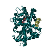
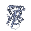
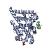
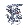
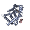
Entry Database : PDB / ID : 2wb9Title Fasciola hepatica sigma class GST GLUTATHIONE TRANSFERASE SIGMA CLASS Keywords / Function / homology Function Domain/homology Component
/ / / / / / / / / / / / / / / / / / / / / / / / / Biological species FASCIOLA HEPATICA (liver fluke)Method / / Resolution : 1.59 Å Authors Line, K. / Isupov, M.N. / LaCourse, J. / Brophy, P.M. / Littlechild, J.A. Journal : To be Published Title : The 1.6 Angstrom Crystal Structure of the Fasciola Hepatica Sigma Class GstAuthors : Line, K. / Isupov, M.N. / Lacourse, J. / Brophy, P.M. / Littlechild, J.A. History Deposition Feb 23, 2009 Deposition site / Processing site Revision 1.0 Mar 31, 2010 Provider / Type Revision 1.1 Feb 1, 2012 Group Atomic model / Derived calculations ... Atomic model / Derived calculations / Non-polymer description / Other / Version format compliance Revision 1.2 Dec 13, 2023 Group Data collection / Database references ... Data collection / Database references / Derived calculations / Other / Refinement description Category chem_comp_atom / chem_comp_bond ... chem_comp_atom / chem_comp_bond / database_2 / pdbx_database_status / pdbx_initial_refinement_model / struct_site Item _database_2.pdbx_DOI / _database_2.pdbx_database_accession ... _database_2.pdbx_DOI / _database_2.pdbx_database_accession / _pdbx_database_status.status_code_sf / _struct_site.pdbx_auth_asym_id / _struct_site.pdbx_auth_comp_id / _struct_site.pdbx_auth_seq_id
Show all Show less
 Movie
Movie Controller


 Open data
 Basic information
 Components
 Keywords
 TRANSFERASE / THIOREDOXIN FOLD
 Function and homology information
 Authors
 Citation
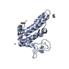
 Structure visualization
 Downloads & links
 Other downloads


 PDBj
PDBj


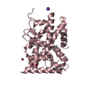
 Assembly
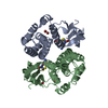


 Mass: 307.323 Da / Num. of mol.: 2 / Source method: obtained synthetically / Formula: C10H17N3O6S
 Type: L-peptide linking / Mass: 121.158 Da / Num. of mol.: 1 / Source method: obtained synthetically / Formula: C3H7NO2S
 Mass: 79.904 Da / Num. of mol.: 5 / Source method: obtained synthetically / Formula: Br
 Mass: 18.015 Da / Num. of mol.: 496 / Source method: isolated from a natural source / Formula: H2O
 Sample preparation
 Processing