 Movie
Movie Controller

[English] 日本語
 Yorodumi
- PDB-2wam: Crystal structure of Mycobacterium tuberculosis unknown function ... -
+ Open data

- Basic information
| Entry | Database: PDB / ID: 2wam | ||||||
|---|---|---|---|---|---|---|---|
| Title | Crystal structure of Mycobacterium tuberculosis unknown function protein Rv2714 | ||||||
 Components Components | CONSERVED HYPOTHETICAL ALANINE AND LEUCINE RICH PROTEIN | ||||||
 Keywords Keywords | UNKNOWN FUNCTION | ||||||
| Function / homology |  Function and homology information Function and homology information | ||||||
| Biological species |    MYCOBACTERIUM TUBERCULOSIS (bacteria) MYCOBACTERIUM TUBERCULOSIS (bacteria) | ||||||
| Method | X-RAY DIFFRACTION / SYNCHROTRON / MOLECULAR REPLACEMENT / Resolution: 2.6 Å | ||||||
 Authors Authors | Bellinzoni, M. / Grana, M. / Buschiazzo, A. / Miras, I. / Haouz, A. / Alzari, P.M. | ||||||
 Citation Citation | Journal: Acta Crystallogr.,Sect.F / Year: 2009 Title: Structure of Mycobacterium Tuberculosis Rv2714, a Representative of a Duplicated Gene Family in Actinobacteria. Authors: Grana, M. / Bellinzoni, M. / Miras, I. / Fiex-Vandal, C. / Haouz, A. / Shepard, W. / Buschiazzo, A. / Alzari, P.M. | ||||||
| History |
|
- Structure visualization
| Structure viewer | Molecule: MolmilJmol/JSmol |
|---|
- Downloads & links
-Download
| PDBx/mmCIF format | 2wam.cif.gz | 342.6 KB | Display | PDBx/mmCIF format |
|---|---|---|---|---|
| PDB format | pdb2wam.ent.gz | 279.8 KB | Display | PDB format |
| PDBx/mmJSON format | 2wam.json.gz | Tree view | PDBx/mmJSON format | |
| Others |  Other downloads Other downloads |
-Validation report
| Arichive directory | https://data.pdbj.org/pub/pdb/validation_reports/wa/2wamftp://data.pdbj.org/pub/pdb/validation_reports/wa/2wam | HTTPS FTP |
|---|
-Related structure data
| Similar structure data |
|---|

-Links
 PDBj
PDBj- Assembly
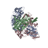
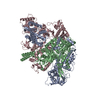
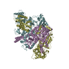
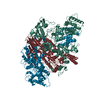
| Deposited unit | 
| ||||||||||||
|---|---|---|---|---|---|---|---|---|---|---|---|---|---|
| 1 | 
| ||||||||||||
| 2 |
| ||||||||||||
| Unit cell |
| ||||||||||||
| Noncrystallographic symmetry (NCS) | NCS oper:
|
-Components
| #1: Protein | Mass: 38817.207 Da / Num. of mol.: 3 / Fragment: RESIDUES 2-323 Source method: isolated from a genetically manipulated source Source: (gene. exp.) MYCOBACTERIUM TUBERCULOSIS (bacteria) / Strain: H37RV / Plasmid: PDEST17 / Production host: ESCHERICHIA COLI (E. coli) / Strain (production host): BL21(DE3)PLYSS / References: UniProt: O07213#2: Chemical | Glycerol  Mass: 92.094 Da / Num. of mol.: 2 / Source method: obtained synthetically / Formula: C3H8O3 Mass: 92.094 Da / Num. of mol.: 2 / Source method: obtained synthetically / Formula: C3H8O3#3: Water | ChemComp-HOH / | Water Mass: 18.015 Da / Num. of mol.: 233 / Source method: isolated from a natural source / Formula: H2O Mass: 18.015 Da / Num. of mol.: 233 / Source method: isolated from a natural source / Formula: H2OSequence details | N-TERMINAL SEQUENCE MSYYHHHHHH | |
|---|
-Experimental details
-Experiment
| Experiment | Method: X-RAY DIFFRACTION / Number of used crystals: 1 |
|---|
- Sample preparation
| Crystal | Density Matthews: 3.18 Å3/Da / Density % sol: 61.3 % / Description: NONE |
|---|---|
| Crystal grow | pH: 6.5 / Details: pH 6.5 |
-Data collection
| Diffraction | Mean temperature: 100 K |
|---|---|
| Diffraction source | Source: SYNCHROTRON / Site: ESRF  / Beamline: ID14-3 / Wavelength: 0.931 / Beamline: ID14-3 / Wavelength: 0.931 |
| Detector | Type: ADSC CCD / Detector: CCD / Date: Jun 20, 2007 |
| Radiation | Protocol: SINGLE WAVELENGTH / Monochromatic (M) / Laue (L): M / Scattering type: x-ray |
| Radiation wavelength | Wavelength: 0.931 Å / Relative weight: 1 |
| Reflection | Resolution: 2.6→47.66 Å / Num. obs: 45795 / % possible obs: 99.8 % / Observed criterion σ(I): 2 / Redundancy: 3.7 % / Biso Wilson estimate: 55.8 Å2 / Rmerge(I) obs: 0.07 / Net I/σ(I): 14.8 |
| Reflection shell | Resolution: 2.6→2.74 Å / Redundancy: 3.8 % / Rmerge(I) obs: 0.53 / Mean I/σ(I) obs: 2.4 / % possible all: 100 |
- Processing
| Software |
| ||||||||||||||||||||||||||||||||||||||||||||||||||||||||||||||||||||||||||||||||||||||||||||||||||||||||||||||||||||||||||||||
|---|---|---|---|---|---|---|---|---|---|---|---|---|---|---|---|---|---|---|---|---|---|---|---|---|---|---|---|---|---|---|---|---|---|---|---|---|---|---|---|---|---|---|---|---|---|---|---|---|---|---|---|---|---|---|---|---|---|---|---|---|---|---|---|---|---|---|---|---|---|---|---|---|---|---|---|---|---|---|---|---|---|---|---|---|---|---|---|---|---|---|---|---|---|---|---|---|---|---|---|---|---|---|---|---|---|---|---|---|---|---|---|---|---|---|---|---|---|---|---|---|---|---|---|---|---|---|---|
| Refinement | Method to determine structure: MOLECULAR REPLACEMENT Starting model: UNPUBLISHED Resolution: 2.6→44.38 Å / SU ML: 0.33 / σ(F): 1.35 / Phase error: 20.31 / Stereochemistry target values: ML Details: HYDROGENS HAVE BEEN ADDED IN THE RIDING POSITIONS. ATOM RECORDS CONTAIN ISOTROPIC EQUIVALENTS OF THE SUM OF TLS AND RESIDUAL B FACTORS. ANISOU RECORDS CONTAIN SUM OF TLS AND RESIDUAL U FACTORS.
| ||||||||||||||||||||||||||||||||||||||||||||||||||||||||||||||||||||||||||||||||||||||||||||||||||||||||||||||||||||||||||||||
| Solvent computation | Shrinkage radii: 0.9 Å / VDW probe radii: 1.11 Å / Solvent model: FLAT BULK SOLVENT MODEL / Bsol: 45.03 Å2 / ksol: 0.34 e/Å3 | ||||||||||||||||||||||||||||||||||||||||||||||||||||||||||||||||||||||||||||||||||||||||||||||||||||||||||||||||||||||||||||||
| Displacement parameters | Biso mean: 50.8 Å2
| ||||||||||||||||||||||||||||||||||||||||||||||||||||||||||||||||||||||||||||||||||||||||||||||||||||||||||||||||||||||||||||||
| Refinement step | Cycle: LAST / Resolution: 2.6→44.38 Å
| ||||||||||||||||||||||||||||||||||||||||||||||||||||||||||||||||||||||||||||||||||||||||||||||||||||||||||||||||||||||||||||||
| Refine LS restraints |
| ||||||||||||||||||||||||||||||||||||||||||||||||||||||||||||||||||||||||||||||||||||||||||||||||||||||||||||||||||||||||||||||
| LS refinement shell |
| ||||||||||||||||||||||||||||||||||||||||||||||||||||||||||||||||||||||||||||||||||||||||||||||||||||||||||||||||||||||||||||||
| Refinement TLS params. | Method: refined / Refine-ID: X-RAY DIFFRACTION
| ||||||||||||||||||||||||||||||||||||||||||||||||||||||||||||||||||||||||||||||||||||||||||||||||||||||||||||||||||||||||||||||
| Refinement TLS group |
|
