 Movie
Movie Controller


+ Open data

- Basic information
| Entry | Database: PDB / ID: 2w9f | ||||||
|---|---|---|---|---|---|---|---|
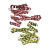
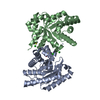
| Title | Crystal Structure of CDK4 in complex with a D-type cyclin | ||||||
 Components Components |
| ||||||
 Keywords Keywords |  CELL CYCLE / SERINE/THREONINE-PROTEIN KINASE / CHROMOSOMAL REARRANGEMENT / ATP-BINDING / TRANSFERASE / POLYMORPHISM / CELL DIVISION / PROTO-ONCOGENE / PHOSPHOPROTEIN / DISEASE MUTATION / NUCLEOTIDE-BINDING / CYCLIN DEPENDENT KINASE / KINASE / CYCLIN / ONCOLOGY / DRUG DESGN CELL CYCLE / SERINE/THREONINE-PROTEIN KINASE / CHROMOSOMAL REARRANGEMENT / ATP-BINDING / TRANSFERASE / POLYMORPHISM / CELL DIVISION / PROTO-ONCOGENE / PHOSPHOPROTEIN / DISEASE MUTATION / NUCLEOTIDE-BINDING / CYCLIN DEPENDENT KINASE / KINASE / CYCLIN / ONCOLOGY / DRUG DESGN | ||||||
| Function / homology |  Function and homology information Function and homology informationre-entry into mitotic cell cycle / cyclin D3-CDK4 complex / cyclin D1-CDK4 complex / cyclin D2-CDK4 complex / Evasion of Oncogene Induced Senescence Due to Defective p16INK4A binding to CDK4 / Evasion of Oxidative Stress Induced Senescence Due to Defective p16INK4A binding to CDK4 / cellular response to ionomycin / regulation of transcription initiation by RNA polymerase II / Drug-mediated inhibition of CDK4/CDK6 activity / Evasion of Oncogene Induced Senescence Due to Defective p16INK4A binding to CDK4 and CDK6 ...re-entry into mitotic cell cycle / cyclin D3-CDK4 complex / cyclin D1-CDK4 complex / cyclin D2-CDK4 complex / Evasion of Oncogene Induced Senescence Due to Defective p16INK4A binding to CDK4 / Evasion of Oxidative Stress Induced Senescence Due to Defective p16INK4A binding to CDK4 / cellular response to ionomycin / regulation of transcription initiation by RNA polymerase II / Drug-mediated inhibition of CDK4/CDK6 activity / Evasion of Oncogene Induced Senescence Due to Defective p16INK4A binding to CDK4 and CDK6 / Evasion of Oxidative Stress Induced Senescence Due to Defective p16INK4A binding to CDK4 and CDK6 / regulation of type B pancreatic cell proliferation / RUNX3 regulates WNT signaling / response to leptin / positive regulation of mammary gland epithelial cell proliferation / Transcriptional regulation by RUNX2 / cellular response to phorbol 13-acetate 12-myristate / mitotic cell cycle phase transition / cyclin-dependent protein serine/threonine kinase activator activity / proline-rich region binding / Regulation of RUNX1 Expression and Activity / cyclin-dependent protein serine/threonine kinase regulator activity / mammary gland epithelial cell proliferation / response to UV-A / negative regulation of epithelial cell differentiation / fat cell differentiation / PTK6 Regulates Cell Cycle / Defective binding of RB1 mutants to E2F1,(E2F2, E2F3) / RUNX3 regulates p14-ARF / positive regulation of cyclin-dependent protein serine/threonine kinase activity / Transcriptional Regulation by VENTX / Estrogen-dependent nuclear events downstream of ESR-membrane signaling / cyclin-dependent protein kinase holoenzyme complex / bicellular tight junction / mammary gland alveolus development / cyclin-dependent kinase / cyclin-dependent protein serine/threonine kinase activity / endoplasmic reticulum unfolded protein response / mitotic G1 DNA damage checkpoint signaling / positive regulation of G2/M transition of mitotic cell cycle / regulation of G2/M transition of mitotic cell cycle / transcription repressor complex / cellular response to interleukin-4 / lactation / cyclin binding / response to organic substance / Ubiquitin-dependent degradation of Cyclin D / liver regeneration / G1/S transition of mitotic cell cycle / Oncogene Induced Senescence / neuron differentiation / SCF(Skp2)-mediated degradation of p27/p21 / RMTs methylate histone arginines / Wnt signaling pathway / Meiotic recombination / Pre-NOTCH Transcription and Translation / Transcriptional regulation of white adipocyte differentiation / Transcriptional regulation of granulopoiesis / histone deacetylase binding / transcription corepressor activity / Cyclin D associated events in G1 / positive regulation of fibroblast proliferation / Senescence-Associated Secretory Phenotype (SASP) / regulation of gene expression / Oxidative Stress Induced Senescence / Interleukin-4 and Interleukin-13 signaling / nuclear membrane / Estrogen-dependent gene expression / cellular response to lipopolysaccharide / negative regulation of neuron apoptotic process / transcription regulator complex / regulation of cell cycle / protein kinase activity / response to xenobiotic stimulus / positive regulation of protein phosphorylation / cell division / protein phosphorylation / protein serine kinase activity / DNA damage response / positive regulation of cell population proliferation / chromatin / nucleolus / protein kinase binding / negative regulation of transcription by RNA polymerase II / enzyme binding / signal transduction / nucleoplasm / ATP binding / nucleus / cytosol / cytoplasmSimilarity search - Function | ||||||
| Biological species |  HOMO SAPIENS (human) HOMO SAPIENS (human) | ||||||
| Method | X-RAY DIFFRACTION / SYNCHROTRON / MOLECULAR REPLACEMENT / Resolution: 2.85 Å | ||||||
 Authors Authors | Day, P.J. / Cleasby, A. / Tickle, I.J. / Reilly, M.O. / Coyle, J.E. / Holding, F.P. / McMenamin, R.L. / Yon, J. / Chopra, R. / Lengauer, C. / Jhoti, H. | ||||||
 Citation Citation | Journal: Proc.Natl.Acad.Sci.USA / Year: 2009 Title: Crystal Structure of Human Cdk4 in Complex with a D-Type Cyclin. Authors: Day, P.J. / Cleasby, A. / Tickle, I.J. / O'Reilly, M. / Coyle, J.E. / Holding, F.P. / Mcmenamin, R.L. / Yon, J. / Chopra, R. / Lengauer, C. / Jhoti, H. | ||||||
| History |
|
- Structure visualization
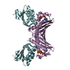
| Structure viewer | Molecule: MolmilJmol/JSmol |
|---|
- Downloads & links
-Download
| PDBx/mmCIF format | 2w9f.cif.gz | 113.8 KB | Display | PDBx/mmCIF format |
|---|---|---|---|---|
| PDB format | pdb2w9f.ent.gz | 87 KB | Display | PDB format |
| PDBx/mmJSON format | 2w9f.json.gz | Tree view | PDBx/mmJSON format | |
| Others |  Other downloads Other downloads |
-Validation report
| Arichive directory | https://data.pdbj.org/pub/pdb/validation_reports/w9/2w9fftp://data.pdbj.org/pub/pdb/validation_reports/w9/2w9f | HTTPS FTP |
|---|
-Related structure data
| Related structure data |  2w96C  2w99C 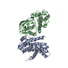 2w9zC 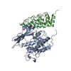 1blxS C: citing same article ( S: Starting model for refinement |
|---|---|
| Similar structure data |


-Links
 PDBj
PDBj


- Assembly
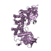
| Deposited unit | 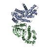
| ||||||||
|---|---|---|---|---|---|---|---|---|---|
| 1 |
| ||||||||
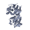
| Unit cell |
|
-Components
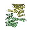
| #1: Protein | Mass: 30990.307 Da / Num. of mol.: 1 Source method: isolated from a genetically manipulated source Source: (gene. exp.) HOMO SAPIENS (human) / Cell line (production host): Sf21 / Production host:   SPODOPTERA FRUGIPERDA (fall armyworm) / References: UniProt: P24385 SPODOPTERA FRUGIPERDA (fall armyworm) / References: UniProt: P24385 |
|---|---|
| #2: Protein | / CDK4 / CYCLIN-DEPENDENT KINASE 4 / PSK-J3 Mass: 34620.723 Da / Num. of mol.: 1 / Fragment: KINASE DOMAIN, RESIDUES 1-44,48-303 / Mutation: YES Source method: isolated from a genetically manipulated source Source: (gene. exp.) HOMO SAPIENS (human) / Cell line (production host): Sf21 / Production host: SPODOPTERA FRUGIPERDA (fall armyworm) / References: UniProt: P11802, cyclin-dependent kinase |
| #3: Water | ChemComp-HOH / Water Mass: 18.015 Da / Num. of mol.: 70 / Source method: isolated from a natural source / Formula: H2O Mass: 18.015 Da / Num. of mol.: 70 / Source method: isolated from a natural source / Formula: H2O |
| Compound details | ENGINEERED RESIDUE IN CHAIN B, GLY 43 TO GLU ENGINEERED RESIDUE IN CHAIN B, GLY 44 TO GLU ...ENGINEERED |
-Experimental details
-Experiment
| Experiment | Method: X-RAY DIFFRACTION / Number of used crystals: 1 |
|---|
- Sample preparation
| Crystal | Density Matthews: 3.09 Å3/Da / Density % sol: 59.94 % / Description: NONE |
|---|
-Data collection
| Diffraction | Mean temperature: 100 K |
|---|---|
| Diffraction source | Source: SYNCHROTRON / Site: ESRF  / Beamline: ID23-1 / Wavelength: 0.931 / Beamline: ID23-1 / Wavelength: 0.931 |
| Detector | Type: ADSC CCD / Detector: CCD / Date: Jun 24, 2007 |
| Radiation | Protocol: SINGLE WAVELENGTH / Monochromatic (M) / Laue (L): M / Scattering type: x-ray |
| Radiation wavelength | Wavelength: 0.931 Å / Relative weight: 1 |
| Reflection | Resolution: 2.85→60.9 Å / Num. obs: 16778 / % possible obs: 98.6 % / Observed criterion σ(I): 0 / Redundancy: 3.1 % / Biso Wilson estimate: 82.07 Å2 / Rmerge(I) obs: 0.08 / Net I/σ(I): 5.7 |
| Reflection shell | Resolution: 2.85→3 Å / Rmerge(I) obs: 0.41 / Mean I/σ(I) obs: 1.9 / % possible all: 99.3 |
- Processing
| Software |
| ||||||||||||||||||||
|---|---|---|---|---|---|---|---|---|---|---|---|---|---|---|---|---|---|---|---|---|---|
| Refinement | Method to determine structure: MOLECULAR REPLACEMENT Starting model: PDB ENTRY 1BLX Resolution: 2.85→93.66 Å / Cross valid method: THROUGHOUT / σ(F): 0 Details: HYDROGENS HAVE BEEN ADDED IN THE RIDING POSITIONS. DISORDERED RESIDUES WERE MODELLED STEREOCHEMICALLY WHEN POSSIBLE
| ||||||||||||||||||||
| Displacement parameters | Biso mean: 84.35 Å2
| ||||||||||||||||||||
| Refinement step | Cycle: LAST / Resolution: 2.85→93.66 Å
| ||||||||||||||||||||
| LS refinement shell | Resolution: 2.85→3.02 Å / Total num. of bins used: 9
|
