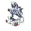
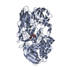
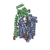
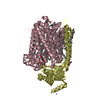
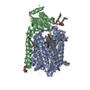
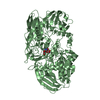
[English] 日本語
Yorodumi
- PDB-2vzv: Substrate Complex of Amycolatopsis orientalis exo-chitosanase Csx... -

+
Open data


ID or keywords:

Loading...

-
Basic information

Entry
Database: PDB / ID: 2vzv
TitleSubstrate Complex of Amycolatopsis orientalis exo-chitosanase CsxA E541A with chitosan
ComponentsEXO-BETA-D-GLUCOSAMINIDASEExo-1,4-beta-D-glucosaminidase
KeywordsHYDROLASE / EXO-BETA-D-GLUCOSAMINIDASE / GH2 / CSXA / CHITOSAN / GLYCOSIDE HYDROLASE
Function / homology
Function and homology information


exo-1,4-beta-D-glucosaminidase / exo-1,4-beta-D-glucosaminidase activity / chitin catabolic process / polysaccharide catabolic process / hydrolase activity, hydrolyzing O-glycosyl compounds / carbohydrate binding / extracellular region
Similarity search - Function
Exo-beta-D-glucosaminidase, Ig-fold domain / Exo-beta-D-glucosaminidase/Endo-beta-mannosidase / Exo-beta-D-glucosaminidase Ig-fold domain / Carbohydrate binding module (family 35) / CBM6 (carbohydrate binding type-6) domain profile. / Carbohydrate binding module family 6 / Glycoside hydrolase, family 2, immunoglobulin-like beta-sandwich / Glycosyl hydrolases family 2, sugar binding domain / Glycosyl hydrolases family 2 / Glycosyl hydrolases family 2, sugar binding domain ...Exo-beta-D-glucosaminidase, Ig-fold domain / Exo-beta-D-glucosaminidase/Endo-beta-mannosidase / Exo-beta-D-glucosaminidase Ig-fold domain / Carbohydrate binding module (family 35) / CBM6 (carbohydrate binding type-6) domain profile. / Carbohydrate binding module family 6 / Glycoside hydrolase, family 2, immunoglobulin-like beta-sandwich / Glycosyl hydrolases family 2, sugar binding domain / Glycosyl hydrolases family 2 / Glycosyl hydrolases family 2, sugar binding domain / Beta-Galactosidase/glucuronidase domain superfamily / Galactose-binding domain-like / Galactose-binding-like domain superfamily / Glycosidases / Glycoside hydrolase superfamily / Jelly Rolls / Immunoglobulins / TIM Barrel / Alpha-Beta Barrel / Immunoglobulin-like fold / Immunoglobulin-like / Sandwich / Mainly Beta / Alpha Beta
Similarity search - Domain/homology
Exo-beta-D-glucosaminidase
Similarity search - Component
Biological speciesAMYCOLATOPSIS ORIENTALIS (bacteria)
MethodX-RAY DIFFRACTION / SYNCHROTRON / OTHER / Resolution: 2.7 Å
AuthorsLammerts van Bueren, A. / Ghinet, M.G. / Gregg, K. / Fleury, A. / Brzezinski, R. / Boraston, A.B.
CitationJournal: J.Mol.Biol. / Year: 2009
Title: The Structural Basis of Substrate Recognition in an Exo-Beta-D-Glucosaminidase Involved in Chitosan Hydrolysis.
Authors: Lammerts Van Bueren, A. / Ghinet, M.G. / Gregg, K. / Fleury, A. / Brzezinski, R. / Boraston, A.B.
History
DepositionAug 5, 2008Deposition site: PDBE / Processing site: PDBE
Revision 1.0Oct 14, 2008Provider: repository / Type: Initial release
Revision 1.1Aug 17, 2011Group: Atomic model / Derived calculations ...Atomic model / Derived calculations / Non-polymer description / Other / Refinement description / Structure summary / Version format compliance
Revision 2.0Jul 29, 2020Group: Advisory / Atomic model ...Advisory / Atomic model / Data collection / Derived calculations / Other / Structure summary
Category: atom_site / chem_comp ...atom_site / chem_comp / entity / pdbx_branch_scheme / pdbx_chem_comp_identifier / pdbx_database_status / pdbx_entity_branch / pdbx_entity_branch_descriptor / pdbx_entity_branch_link / pdbx_entity_branch_list / pdbx_entity_nonpoly / pdbx_nonpoly_scheme / pdbx_struct_assembly_gen / pdbx_unobs_or_zero_occ_atoms / struct_asym / struct_conn / struct_site / struct_site_gen
Item: _atom_site.B_iso_or_equiv / _atom_site.Cartn_x ..._atom_site.B_iso_or_equiv / _atom_site.Cartn_x / _atom_site.Cartn_y / _atom_site.Cartn_z / _atom_site.auth_asym_id / _atom_site.auth_atom_id / _atom_site.auth_seq_id / _atom_site.label_asym_id / _atom_site.label_atom_id / _atom_site.type_symbol / _chem_comp.name / _chem_comp.type / _entity.formula_weight / _entity.pdbx_description / _entity.pdbx_number_of_molecules / _entity.type / _pdbx_database_status.status_code_sf / _pdbx_struct_assembly_gen.asym_id_list / _struct_conn.pdbx_leaving_atom_flag / _struct_conn.ptnr1_auth_asym_id / _struct_conn.ptnr1_auth_seq_id / _struct_conn.ptnr1_label_asym_id / _struct_conn.ptnr1_label_atom_id / _struct_conn.ptnr2_auth_asym_id / _struct_conn.ptnr2_auth_seq_id / _struct_conn.ptnr2_label_asym_id / _struct_conn.ptnr2_label_atom_id
Description: Carbohydrate remediation / Provider: repository / Type: Remediation
Remark 700 SHEET THE SHEET STRUCTURE OF THIS MOLECULE IS BIFURCATED. IN ORDER TO REPRESENT THIS FEATURE IN ... SHEET THE SHEET STRUCTURE OF THIS MOLECULE IS BIFURCATED. IN ORDER TO REPRESENT THIS FEATURE IN THE SHEET RECORDS BELOW, TWO SHEETS ARE DEFINED.

-
Structure visualization

Structure viewerMolecule:
MolmilJmol/JSmol

Downloads & links

-
Assembly

Deposited unit
A: EXO-BETA-D-GLUCOSAMINIDASE
B: EXO-BETA-D-GLUCOSAMINIDASE
hetero molecules


Theoretical massNumber of molelcules
Total (without water)221,8324
Polymers221,1522
Non-polymers6812
Water7,620423
1
A: EXO-BETA-D-GLUCOSAMINIDASE
hetero molecules


Theoretical massNumber of molelcules
Total (without water)110,9162
Polymers110,5761
Non-polymers3401
Water181
TypeNameSymmetry operationNumber
identity operation1_555x,y,z1
MethodPISA
2
B: EXO-BETA-D-GLUCOSAMINIDASE
hetero molecules


Theoretical massNumber of molelcules
Total (without water)110,9162
Polymers110,5761
Non-polymers3401
Water181
TypeNameSymmetry operationNumber
identity operation1_555x,y,z1
MethodPISA
Unit cell
Length a, b, c (Å)86.860, 121.410, 184.710
Angle α, β, γ (deg.)90.00, 90.00, 90.00
Int Tables number19
Space group name H-MP212121

-
Components

#1: Protein EXO-BETA-D-GLUCOSAMINIDASE / Exo-1,4-beta-D-glucosaminidase / CSXA


Mass: 110575.758 Da / Num. of mol.: 2 / Fragment: RESIDUES 2-1032 / Mutation: YES
Source method: isolated from a genetically manipulated source
Source: (gene. exp.) AMYCOLATOPSIS ORIENTALIS (bacteria) / Plasmid: PFD666 / Production host: STREPTOMYCES LIVIDANS (bacteria) / Strain (production host): TK24 / References: UniProt: Q56F26
#2: Polysaccharide 2-amino-2-deoxy-beta-D-glucopyranose-(1-4)-2-amino-2-deoxy-beta-D-glucopyranose


Type: oligosaccharide / Mass: 340.327 Da / Num. of mol.: 2
Source method: isolated from a genetically manipulated source
DescriptorTypeProgram
DGlcpNb1-4DGlcpNb1-ROHGlycam Condensed SequenceGMML 1.0
WURCS=2.0/1,2,1/[a2122h-1b_1-5_2*N]/1-1/a4-b1WURCSPDB2Glycan 1.1.0
[][b-D-GlcpN]{[(4+1)][b-D-GlcpN]{}}LINUCSPDB-CARE
#3: Water ChemComp-HOH / water / Water


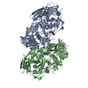
Mass: 18.015 Da / Num. of mol.: 423 / Source method: isolated from a natural source / Formula: H2O
Compound detailsENGINEERED RESIDUE IN CHAIN A, GLU 541 TO ALA ENGINEERED RESIDUE IN CHAIN B, GLU 541 TO ALA

-
Experimental details

-
Experiment

ExperimentMethod: X-RAY DIFFRACTION

-
Sample preparation

CrystalDensity Matthews: 2.56 Å3/Da / Density % sol: 51.66 % / Description: NONE

-
Data collection

DiffractionMean temperature: 113 K
Diffraction sourceSource: SYNCHROTRON / Site: NSLS / Beamline: X8C / Wavelength: 1.1
DetectorType: ADSC CCD / Detector: CCD
RadiationProtocol: SINGLE WAVELENGTH / Monochromatic (M) / Laue (L): M / Scattering type: x-ray
Radiation wavelengthWavelength: 1.1 Å / Relative weight: 1
ReflectionResolution: 2.7→20 Å / Num. obs: 47221 / % possible obs: 91.8 % / Observed criterion σ(I): 2 / Redundancy: 7.7 % / Rmerge(I) obs: 0.12 / Net I/σ(I): 15.4

-
Processing

SoftwareName: REFMAC / Version: 5.2.0019 / Classification: refinement
RefinementMethod to determine structure: OTHER / Resolution: 2.7→185.7 Å / Cor.coef. Fo:Fc: 0.923 / Cor.coef. Fo:Fc free: 0.853 / SU B: 12.465 / SU ML: 0.257 / Cross valid method: THROUGHOUT / ESU R Free: 0.387 / Stereochemistry target values: MAXIMUM LIKELIHOOD / Details: HYDROGENS HAVE BEEN ADDED IN THE RIDING POSITIONS.
RfactorNum. reflection% reflectionSelection details
Rfree0.255 2549 5.1 %RANDOM
Rwork0.182 ---
obs0.186 47221 91.3 %-
Solvent computationIon probe radii: 0.8 Å / Shrinkage radii: 0.8 Å / VDW probe radii: 1.2 Å / Solvent model: MASK
Displacement parametersBiso mean: 15.58 Å2
Baniso -1Baniso -2Baniso -3
1-0.01 Å20 Å20 Å2
2---0.58 Å20 Å2
3---0.58 Å2
Refinement stepCycle: LAST / Resolution: 2.7→185.7 Å
ProteinNucleic acidLigandSolventTotal
Num. atoms13096 0 46 423 13565
Refine LS restraints
Refine-IDTypeDev idealDev ideal targetNumber
X-RAY DIFFRACTIONr_bond_refined_d0.010.02213464
X-RAY DIFFRACTIONr_bond_other_d
X-RAY DIFFRACTIONr_angle_refined_deg1.2821.94518380
X-RAY DIFFRACTIONr_angle_other_deg
X-RAY DIFFRACTIONr_dihedral_angle_1_deg6.12951712
X-RAY DIFFRACTIONr_dihedral_angle_2_deg34.70824.271576
X-RAY DIFFRACTIONr_dihedral_angle_3_deg16.117152050
X-RAY DIFFRACTIONr_dihedral_angle_4_deg19.6241570
X-RAY DIFFRACTIONr_chiral_restr0.0780.22038
X-RAY DIFFRACTIONr_gen_planes_refined0.0040.0210350
X-RAY DIFFRACTIONr_gen_planes_other
X-RAY DIFFRACTIONr_nbd_refined0.2050.26353
X-RAY DIFFRACTIONr_nbd_other
X-RAY DIFFRACTIONr_nbtor_refined0.3060.29034
X-RAY DIFFRACTIONr_nbtor_other
X-RAY DIFFRACTIONr_xyhbond_nbd_refined0.1510.2763
X-RAY DIFFRACTIONr_xyhbond_nbd_other
X-RAY DIFFRACTIONr_metal_ion_refined
X-RAY DIFFRACTIONr_metal_ion_other
X-RAY DIFFRACTIONr_symmetry_vdw_refined0.6010.262
X-RAY DIFFRACTIONr_symmetry_vdw_other
X-RAY DIFFRACTIONr_symmetry_hbond_refined0.2840.211
X-RAY DIFFRACTIONr_symmetry_hbond_other
X-RAY DIFFRACTIONr_symmetry_metal_ion_refined
X-RAY DIFFRACTIONr_symmetry_metal_ion_other
X-RAY DIFFRACTIONr_mcbond_it0.3981.58658
X-RAY DIFFRACTIONr_mcbond_other
X-RAY DIFFRACTIONr_mcangle_it0.707213696
X-RAY DIFFRACTIONr_mcangle_other
X-RAY DIFFRACTIONr_scbond_it0.92735557
X-RAY DIFFRACTIONr_scbond_other
X-RAY DIFFRACTIONr_scangle_it1.5054.54684
X-RAY DIFFRACTIONr_scangle_other
X-RAY DIFFRACTIONr_long_range_B_refined
X-RAY DIFFRACTIONr_long_range_B_other
X-RAY DIFFRACTIONr_rigid_bond_restr
X-RAY DIFFRACTIONr_sphericity_free
X-RAY DIFFRACTIONr_sphericity_bonded
LS refinement shellResolution: 2.7→2.77 Å / Total num. of bins used: 20 /
RfactorNum. reflection
Rfree0.312 166
Rwork0.227 2934

+
About Yorodumi

-
News

-
Feb 9, 2022. New format data for meta-information of EMDB entries

New format data for meta-information of EMDB entries

  • Version 3 of the EMDB header file is now the official format.
  • The previous official version 1.9 will be removed from the archive.

Related info.:EMDB header

External links:wwPDB to switch to version 3 of the EMDB data model

-
Aug 12, 2020. Covid-19 info

Covid-19 info

URL: https://pdbj.org/emnavi/covid19.php

New page: Covid-19 featured information page in EM Navigator.

Related info.:Covid-19 info / Mar 5, 2020. Novel coronavirus structure data

+
Mar 5, 2020. Novel coronavirus structure data

Novel coronavirus structure data

Related info.:Yorodumi Speices / Aug 12, 2020. Covid-19 info

External links:COVID-19 featured content - PDBj / Molecule of the Month (242):Coronavirus Proteases

+
Jan 31, 2019. EMDB accession codes are about to change! (news from PDBe EMDB page)

EMDB accession codes are about to change! (news from PDBe EMDB page)

  • The allocation of 4 digits for EMDB accession codes will soon come to an end. Whilst these codes will remain in use, new EMDB accession codes will include an additional digit and will expand incrementally as the available range of codes is exhausted. The current 4-digit format prefixed with “EMD-” (i.e. EMD-XXXX) will advance to a 5-digit format (i.e. EMD-XXXXX), and so on. It is currently estimated that the 4-digit codes will be depleted around Spring 2019, at which point the 5-digit format will come into force.
  • The EM Navigator/Yorodumi systems omit the EMD- prefix.

Related info.:Q: What is EMD? / ID/Accession-code notation in Yorodumi/EM Navigator

External links:EMDB Accession Codes are Changing Soon! / Contact to PDBj

+
Jul 12, 2017. Major update of PDB

Major update of PDB

  • wwPDB released updated PDB data conforming to the new PDBx/mmCIF dictionary.
  • This is a major update changing the version number from 4 to 5, and with Remediation, in which all the entries are updated.
  • In this update, many items about electron microscopy experimental information are reorganized (e.g. em_software).
  • Now, EM Navigator and Yorodumi are based on the updated data.

External links:wwPDB Remediation / Enriched Model Files Conforming to OneDep Data Standards Now Available in the PDB FTP Archive

-
Yorodumi

Thousand views of thousand structures

  • Yorodumi is a browser for structure data from EMDB, PDB, SASBDB, etc.
  • This page is also the successor to EM Navigator detail page, and also detail information page/front-end page for Omokage search.
  • The word "yorodu" (or yorozu) is an old Japanese word meaning "ten thousand". "mi" (miru) is to see.

Related info.:EMDB / PDB / SASBDB / Comparison of 3 databanks / Yorodumi Search / Aug 31, 2016. New EM Navigator & Yorodumi / Yorodumi Papers / Jmol/JSmol / Function and homology information / Changes in new EM Navigator and Yorodumi

Read more