 Movie
Movie Controller


+ Open data

- Basic information
| Entry | Database: PDB / ID: 2vyi | ||||||
|---|---|---|---|---|---|---|---|
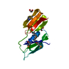
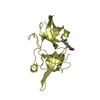
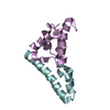
| Title | Crystal Structure of the TPR domain of Human SGT | ||||||
 Components Components | SGTA PROTEIN | ||||||
 Keywords Keywords | CHAPERONE / SGT / TPR REPEAT / PHOSPHOPROTEIN / TETRATRICOPEPTIDE REPEAT PROTEIN / HOST-VIRUS INTERACTION | ||||||
| Function / homology |  Function and homology information Function and homology informationpositive regulation of chaperone-mediated protein folding / : / : / TRC complex / positive regulation of ERAD pathway / tail-anchored membrane protein insertion into ER membrane / extrinsic component of synaptic vesicle membrane / post-translational protein targeting to endoplasmic reticulum membrane / positive regulation of ubiquitin-dependent protein catabolic process / BAT3 complex binding ...positive regulation of chaperone-mediated protein folding / : / : / TRC complex / positive regulation of ERAD pathway / tail-anchored membrane protein insertion into ER membrane / extrinsic component of synaptic vesicle membrane / post-translational protein targeting to endoplasmic reticulum membrane / positive regulation of ubiquitin-dependent protein catabolic process / BAT3 complex binding / Insertion of tail-anchored proteins into the endoplasmic reticulum membrane / : / viral process / negative regulation of ubiquitin-dependent protein catabolic process / : / molecular adaptor activity / membrane / identical protein binding / nucleus / cytosol / cytoplasmSimilarity search - Function | ||||||
| Biological species |  HOMO SAPIENS (human) HOMO SAPIENS (human) | ||||||
| Method | X-RAY DIFFRACTION / SYNCHROTRON / MOLECULAR REPLACEMENT / Resolution: 2.4 Å | ||||||
 Authors Authors | Dutta, S. / Tan, Y.J. | ||||||
 Citation Citation | Journal: Biochemistry / Year: 2008 Title: Structural and Functional Characterization of Human Sgt and its Interaction with Vpu of the Human Immunodeficiency Virus Type 1. Authors: Dutta, S. / Tan, Y.J. | ||||||
| History |
|
- Structure visualization
| Structure viewer | Molecule: MolmilJmol/JSmol |
|---|
- Downloads & links
-Download
| PDBx/mmCIF format | 2vyi.cif.gz | 63.5 KB | Display | PDBx/mmCIF format |
|---|---|---|---|---|
| PDB format | pdb2vyi.ent.gz | 47 KB | Display | PDB format |
| PDBx/mmJSON format | 2vyi.json.gz | Tree view | PDBx/mmJSON format | |
| Others |  Other downloads Other downloads |
-Validation report
| Arichive directory | https://data.pdbj.org/pub/pdb/validation_reports/vy/2vyiftp://data.pdbj.org/pub/pdb/validation_reports/vy/2vyi | HTTPS FTP |
|---|
-Related structure data
| Related structure data |  1waoS S: Starting model for refinement |
|---|---|
| Similar structure data |


-Links
 PDBj
PDBj


- Assembly
| Deposited unit | 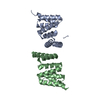
| ||||||||
|---|---|---|---|---|---|---|---|---|---|
| 1 | 
| ||||||||
| 2 | 
| ||||||||
| Unit cell |
|
-Components
| #1: Protein | / SMALL GLUTAMINE RICH PROTEIN WITH TETRATRICOPEPTIDE REPEAT 1 / SMALL GLUTAMINE-RICH ...SMALL GLUTAMINE RICH PROTEIN WITH TETRATRICOPEPTIDE REPEAT 1 / SMALL GLUTAMINE-RICH TETRATRICOPEPTIDE REPEAT (TPR)-CONTAINING / ALPHA / ISOFORM CRA_A Mass: 14430.173 Da / Num. of mol.: 2 / Fragment: TETRATRICOPEPTIDE REPEAT, RESIDUES 84-210 Source method: isolated from a genetically manipulated source Source: (gene. exp.) HOMO SAPIENS (human) / Plasmid: PGEX6P1 / Production host:  ESCHERICHIA COLI (E. coli) / Strain (production host): BL21 / References: UniProt: Q6FIA9, UniProt: O43765*PLUS ESCHERICHIA COLI (E. coli) / Strain (production host): BL21 / References: UniProt: Q6FIA9, UniProt: O43765*PLUS#2: Water | ChemComp-HOH / | Water Mass: 18.015 Da / Num. of mol.: 180 / Source method: isolated from a natural source / Formula: H2O Mass: 18.015 Da / Num. of mol.: 180 / Source method: isolated from a natural source / Formula: H2O |
|---|
-Experimental details
-Experiment
| Experiment | Method: X-RAY DIFFRACTION / Number of used crystals: 1 |
|---|
- Sample preparation
| Crystal | Density Matthews: 2.7 Å3/Da / Density % sol: 54.4 % / Description: NONE |
|---|---|
| Crystal grow | Details: 4M SODIUM FORMATE |
-Data collection
| Diffraction | Mean temperature: 100 K |
|---|---|
| Diffraction source | Source: SYNCHROTRON / Site: ESRF  / Beamline: ID14-1 / Wavelength: 0.95 / Beamline: ID14-1 / Wavelength: 0.95 |
| Detector | Type: ADSC CCD / Detector: CCD / Date: Oct 7, 2007 |
| Radiation | Protocol: SINGLE WAVELENGTH / Monochromatic (M) / Laue (L): M / Scattering type: x-ray |
| Radiation wavelength | Wavelength: 0.95 Å / Relative weight: 1 |
| Reflection | Resolution: 2.4→30 Å / Num. obs: 12726 / % possible obs: 100 % / Observed criterion σ(I): 3 / Redundancy: 4.8 % / Biso Wilson estimate: 31.2 Å2 / Rmerge(I) obs: 0.07 / Net I/σ(I): 19.2 |
| Reflection shell | Resolution: 2.4→2.49 Å / Redundancy: 4.7 % / Rmerge(I) obs: 0.22 / Mean I/σ(I) obs: 6.8 / % possible all: 99 |
- Processing
| Software |
| ||||||||||||||||||||||||||||||||||||||||||||||||||||||||||||||||||||||||||||||||
|---|---|---|---|---|---|---|---|---|---|---|---|---|---|---|---|---|---|---|---|---|---|---|---|---|---|---|---|---|---|---|---|---|---|---|---|---|---|---|---|---|---|---|---|---|---|---|---|---|---|---|---|---|---|---|---|---|---|---|---|---|---|---|---|---|---|---|---|---|---|---|---|---|---|---|---|---|---|---|---|---|---|
| Refinement | Method to determine structure: MOLECULAR REPLACEMENT Starting model: PDB ENTRY 1WAO Resolution: 2.4→29.71 Å / Rfactor Rfree error: 0.01 / Data cutoff high absF: 1088998.47 / Isotropic thermal model: RESTRAINED / Cross valid method: THROUGHOUT / σ(F): 0 / Details: BULK SOLVENT MODEL USED
| ||||||||||||||||||||||||||||||||||||||||||||||||||||||||||||||||||||||||||||||||
| Solvent computation | Solvent model: FLAT MODEL / Bsol: 46.7274 Å2 / ksol: 0.4 e/Å3 | ||||||||||||||||||||||||||||||||||||||||||||||||||||||||||||||||||||||||||||||||
| Displacement parameters | Biso mean: 30.8 Å2
| ||||||||||||||||||||||||||||||||||||||||||||||||||||||||||||||||||||||||||||||||
| Refine analyze |
| ||||||||||||||||||||||||||||||||||||||||||||||||||||||||||||||||||||||||||||||||
| Refinement step | Cycle: LAST / Resolution: 2.4→29.71 Å
| ||||||||||||||||||||||||||||||||||||||||||||||||||||||||||||||||||||||||||||||||
| Refine LS restraints |
| ||||||||||||||||||||||||||||||||||||||||||||||||||||||||||||||||||||||||||||||||
| LS refinement shell | Resolution: 2.4→2.55 Å / Rfactor Rfree error: 0.035 / Total num. of bins used: 6
| ||||||||||||||||||||||||||||||||||||||||||||||||||||||||||||||||||||||||||||||||
| Xplor file |
|
