 Movie
Movie Controller

[English] 日本語
 Yorodumi
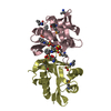
- PDB-2vxk: Structural comparison between Aspergillus fumigatus and human GNA1 -
+ Open data

- Basic information
| Entry | Database: PDB / ID: 2vxk | ||||||
|---|---|---|---|---|---|---|---|
| Title | Structural comparison between Aspergillus fumigatus and human GNA1 | ||||||
 Components Components | GLUCOSAMINE 6-PHOSPHATE ACETYLTRANSFERASE | ||||||
 Keywords Keywords |  TRANSFERASE / KINETICS / UDP-GLCNAC / INHIBITOR DESIGN TRANSFERASE / KINETICS / UDP-GLCNAC / INHIBITOR DESIGN | ||||||
| Function / homology |  Function and homology informationglucosamine-phosphate N-acetyltransferase / glucosamine 6-phosphate N-acetyltransferase activity / UDP-N-acetylglucosamine biosynthetic process / monosaccharide binding / endoplasmic reticulum-Golgi intermediate compartment / Golgi apparatus / endoplasmic reticulum Function and homology informationglucosamine-phosphate N-acetyltransferase / glucosamine 6-phosphate N-acetyltransferase activity / UDP-N-acetylglucosamine biosynthetic process / monosaccharide binding / endoplasmic reticulum-Golgi intermediate compartment / Golgi apparatus / endoplasmic reticulumSimilarity search - Function | ||||||
| Biological species |  ASPERGILLUS FUMIGATUS (mold) ASPERGILLUS FUMIGATUS (mold) | ||||||
| Method | X-RAY DIFFRACTION / SYNCHROTRON / SAD / Resolution: 1.8 Å | ||||||
 Authors Authors | Hurtado-Guerrero, R. / Raimi, O.G. / Min, J. / Zeng, H. / Vallius, L. / Shepherd, S. / Ibrahim, A.F.M. / Wu, H. / Plotnikov, A.N. / van Aalten, D.M.F. | ||||||
 Citation Citation | Journal: Biochem.J. / Year: 2008 Title: Structural and Kinetic Differences between Human and Aspergillus Fumigatus D-Glucosamine-6- Phosphate N-Acetyltransferase. Authors: Hurtado-Guerrero, R. / Raimi, O.G. / Min, J. / Zeng, H. / Vallius, L. / Shepherd, S. / Ibrahim, A.F.M. / Wu, H. / Plotnikov, A.N. / Van Aalten, D.M.F. | ||||||
| History |
|
- Structure visualization
| Structure viewer | Molecule: MolmilJmol/JSmol |
|---|
- Downloads & links
-Download
| PDBx/mmCIF format | 2vxk.cif.gz | 53.6 KB | Display | PDBx/mmCIF format |
|---|---|---|---|---|
| PDB format | pdb2vxk.ent.gz | 38.3 KB | Display | PDB format |
| PDBx/mmJSON format | 2vxk.json.gz | Tree view | PDBx/mmJSON format | |
| Others |  Other downloads Other downloads |
-Validation report
| Arichive directory | https://data.pdbj.org/pub/pdb/validation_reports/vx/2vxkftp://data.pdbj.org/pub/pdb/validation_reports/vx/2vxk | HTTPS FTP |
|---|
-Related structure data
| Related structure data | |
|---|---|
| Similar structure data |


-Links
 PDBj
PDBj


- Assembly
| Deposited unit | 
| ||||||||
|---|---|---|---|---|---|---|---|---|---|
| 1 | 
| ||||||||
| Unit cell |
|
-Components
| #1: Protein | Mass: 21126.113 Da / Num. of mol.: 1 Source method: isolated from a genetically manipulated source Source: (gene. exp.) ASPERGILLUS FUMIGATUS (mold) / Plasmid: PGEX6P1 / Production host:  ESCHERICHIA COLI (E. coli) / Strain (production host): BL21(DE3)PLYSS ESCHERICHIA COLI (E. coli) / Strain (production host): BL21(DE3)PLYSSReferences: UniProt: Q4WCU5, glucosamine-phosphate N-acetyltransferase |
|---|---|
| #2: Sugar | ChemComp-16G / 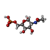  Type: D-saccharide, alpha linking / Mass: 301.188 Da / Num. of mol.: 1 Type: D-saccharide, alpha linking / Mass: 301.188 Da / Num. of mol.: 1Source method: isolated from a genetically manipulated source Formula: C8H16NO9P |
| #3: Chemical | ChemComp-PO4 / Phosphate  Mass: 94.971 Da / Num. of mol.: 1 / Source method: obtained synthetically / Formula: PO4 Mass: 94.971 Da / Num. of mol.: 1 / Source method: obtained synthetically / Formula: PO4 |
| #4: Chemical | ChemComp-COA / Coenzyme A  Mass: 767.534 Da / Num. of mol.: 1 / Source method: obtained synthetically / Formula: C21H36N7O16P3S Mass: 767.534 Da / Num. of mol.: 1 / Source method: obtained synthetically / Formula: C21H36N7O16P3S |
| #5: Water | ChemComp-HOH / Water Mass: 18.015 Da / Num. of mol.: 144 / Source method: isolated from a natural source / Formula: H2O Mass: 18.015 Da / Num. of mol.: 144 / Source method: isolated from a natural source / Formula: H2O |
-Experimental details
-Experiment
| Experiment | Method: X-RAY DIFFRACTION |
|---|
- Sample preparation
| Crystal | Density Matthews: 2.19 Å3/Da / Density % sol: 43.27 % / Description: NONE |
|---|
-Data collection
| Diffraction | Mean temperature: 10 K |
|---|---|
| Diffraction source | Source: SYNCHROTRON / Site: ESRF  / Beamline: BM14 / Wavelength: 0.95 / Beamline: BM14 / Wavelength: 0.95 |
| Detector | Type: MARRESEARCH / Detector: CCD |
| Radiation | Protocol: SINGLE WAVELENGTH / Monochromatic (M) / Laue (L): M / Scattering type: x-ray |
| Radiation wavelength | Wavelength: 0.95 Å / Relative weight: 1 |
| Reflection | Resolution: 1.8→20 Å / Num. obs: 18376 / % possible obs: 98.2 % / Redundancy: 4.6 % / Rmerge(I) obs: 0.03 / Net I/σ(I): 21.2 |
| Reflection shell | Resolution: 1.8→1.86 Å / Redundancy: 4.6 % / Rmerge(I) obs: 0.15 / Mean I/σ(I) obs: 10.5 / % possible all: 98 |
- Processing
| Software | Name: REFMAC / Version: 5.2.0005 / Classification: refinement | ||||||||||||||||||||||||||||||||||||||||||||||||||||||||||||||||||||||||||||||||||||||||||||||||||||||||||||||||||||||||||||||||||||||||||||||||||||||||||||||||||||||||||||||||||||||
|---|---|---|---|---|---|---|---|---|---|---|---|---|---|---|---|---|---|---|---|---|---|---|---|---|---|---|---|---|---|---|---|---|---|---|---|---|---|---|---|---|---|---|---|---|---|---|---|---|---|---|---|---|---|---|---|---|---|---|---|---|---|---|---|---|---|---|---|---|---|---|---|---|---|---|---|---|---|---|---|---|---|---|---|---|---|---|---|---|---|---|---|---|---|---|---|---|---|---|---|---|---|---|---|---|---|---|---|---|---|---|---|---|---|---|---|---|---|---|---|---|---|---|---|---|---|---|---|---|---|---|---|---|---|---|---|---|---|---|---|---|---|---|---|---|---|---|---|---|---|---|---|---|---|---|---|---|---|---|---|---|---|---|---|---|---|---|---|---|---|---|---|---|---|---|---|---|---|---|---|---|---|---|---|
| Refinement | Method to determine structure: SAD Starting model: NONE Resolution: 1.8→20 Å / Cor.coef. Fo:Fc: 0.957 / Cor.coef. Fo:Fc free: 0.942 / SU B: 5.622 / SU ML: 0.081 / TLS residual ADP flag: UNVERIFIED / Cross valid method: THROUGHOUT / ESU R: 0.124 / ESU R Free: 0.121 / Stereochemistry target values: MAXIMUM LIKELIHOOD Details: HYDROGENS HAVE BEEN ADDED IN THE RIDING POSITIONS. COA IS NOT COMPLETE MODELLED SINCE AT THE END OF THE MOLECULE (REGION OF THE THIOL GROUP) WAS COMPLETELY DISORDERED.
| ||||||||||||||||||||||||||||||||||||||||||||||||||||||||||||||||||||||||||||||||||||||||||||||||||||||||||||||||||||||||||||||||||||||||||||||||||||||||||||||||||||||||||||||||||||||
| Solvent computation | Ion probe radii: 0.8 Å / Shrinkage radii: 0.8 Å / VDW probe radii: 1.2 Å / Solvent model: MASK | ||||||||||||||||||||||||||||||||||||||||||||||||||||||||||||||||||||||||||||||||||||||||||||||||||||||||||||||||||||||||||||||||||||||||||||||||||||||||||||||||||||||||||||||||||||||
| Displacement parameters | Biso mean: 12.1 Å2
| ||||||||||||||||||||||||||||||||||||||||||||||||||||||||||||||||||||||||||||||||||||||||||||||||||||||||||||||||||||||||||||||||||||||||||||||||||||||||||||||||||||||||||||||||||||||
| Refinement step | Cycle: LAST / Resolution: 1.8→20 Å
| ||||||||||||||||||||||||||||||||||||||||||||||||||||||||||||||||||||||||||||||||||||||||||||||||||||||||||||||||||||||||||||||||||||||||||||||||||||||||||||||||||||||||||||||||||||||
| Refine LS restraints |
|