 Movie
Movie Controller


+ Open data

- Basic information
| Entry | Database: PDB / ID: 2vul | ||||||
|---|---|---|---|---|---|---|---|
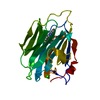
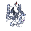
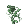
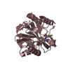
| Title | Thermostable mutant of ENVIRONMENTALLY ISOLATED GH11 XYLANASE | ||||||
 Components Components | GH11 XYLANASE | ||||||
 Keywords Keywords |  HYDROLASE / GH11 / XYLANASE / GLYCOSIDASE HYDROLASE / GH11 / XYLANASE / GLYCOSIDASE | ||||||
| Function / homology |  Function and homology informationendo-1,4-beta-xylanase activity / endo-1,4-beta-xylanase / xylan catabolic process Function and homology informationendo-1,4-beta-xylanase activity / endo-1,4-beta-xylanase / xylan catabolic processSimilarity search - Function | ||||||
| Biological species |  ESCHERICHIA COLI (E. coli) ESCHERICHIA COLI (E. coli) | ||||||
| Method | X-RAY DIFFRACTION / MOLECULAR REPLACEMENT / Resolution: 1.9 Å | ||||||
 Authors Authors | Dumon, C. / Varvak, A. / Wall, M.A. / Flint, J.E. / Lewis, R.J. / Lakey, J.H. / Luginbuhl, P. / Healey, S. / Todaro, T. / Desantis, G. ...Dumon, C. / Varvak, A. / Wall, M.A. / Flint, J.E. / Lewis, R.J. / Lakey, J.H. / Luginbuhl, P. / Healey, S. / Todaro, T. / Desantis, G. / Sun, M. / Parra-Gessert, L. / Tan, X. / Weiner, D.P. / Gilbert, H.J. | ||||||
 Citation Citation | Journal: J.Biol.Chem. / Year: 2008 Title: Engineering Hyperthermostability Into a Gh11 Xylanase is Mediated by Subtle Changes to Protein Structure. Authors: Dumon, C. / Varvak, A. / Wall, M.A. / Flint, J.E. / Lewis, R.J. / Lakey, J.H. / Morland, C. / Luginbuhl, P. / Healey, S. / Todaro, T. / Desantis, G. / Sun, M. / Parra-Gessert, L. / Tan, X. / ...Authors: Dumon, C. / Varvak, A. / Wall, M.A. / Flint, J.E. / Lewis, R.J. / Lakey, J.H. / Morland, C. / Luginbuhl, P. / Healey, S. / Todaro, T. / Desantis, G. / Sun, M. / Parra-Gessert, L. / Tan, X. / Weiner, D.P. / Gilbert, H.J. | ||||||
| History |
|
- Structure visualization
| Structure viewer | Molecule: MolmilJmol/JSmol |
|---|
- Downloads & links
-Download
| PDBx/mmCIF format | 2vul.cif.gz | 60.4 KB | Display | PDBx/mmCIF format |
|---|---|---|---|---|
| PDB format | pdb2vul.ent.gz | 41.2 KB | Display | PDB format |
| PDBx/mmJSON format | 2vul.json.gz | Tree view | PDBx/mmJSON format | |
| Others |  Other downloads Other downloads |
-Validation report
| Arichive directory | https://data.pdbj.org/pub/pdb/validation_reports/vu/2vulftp://data.pdbj.org/pub/pdb/validation_reports/vu/2vul | HTTPS FTP |
|---|
-Related structure data
| Related structure data |  2vujSC S: Starting model for refinement C: citing same article ( |
|---|---|
| Similar structure data |


-Links
 PDBj
PDBj
- Assembly
| Deposited unit | 
| ||||||||
|---|---|---|---|---|---|---|---|---|---|
| 1 |
| ||||||||
| Unit cell |
| ||||||||
| Components on special symmetry positions |
|
-Components
| #1: Protein | Mass: 23437.787 Da / Num. of mol.: 1 / Mutation: YES Source method: isolated from a genetically manipulated source Source: (gene. exp.) ESCHERICHIA COLI (E. coli) / Plasmid: PSE420-CHIS / Production host: ESCHERICHIA COLI (E. coli) / Strain (production host): BL21 / References: UniProt: B2LWN3*PLUS, endo-1,4-beta-xylanase | ||||||
|---|---|---|---|---|---|---|---|
| #2: Chemical | Sulfate  Mass: 96.063 Da / Num. of mol.: 2 / Source method: obtained synthetically / Formula: SO4 Mass: 96.063 Da / Num. of mol.: 2 / Source method: obtained synthetically / Formula: SO4#3: Chemical | Polyethylene glycol  Mass: 546.646 Da / Num. of mol.: 3 / Source method: obtained synthetically / Formula: C24H50O13 / Comment: precipitant*YM Mass: 546.646 Da / Num. of mol.: 3 / Source method: obtained synthetically / Formula: C24H50O13 / Comment: precipitant*YM#4: Water | ChemComp-HOH / | Water Mass: 18.015 Da / Num. of mol.: 192 / Source method: isolated from a natural source / Formula: H2O Mass: 18.015 Da / Num. of mol.: 192 / Source method: isolated from a natural source / Formula: H2OSequence details | THERMOSTABLE MUTATED PROTEIN AN UNIPROT ID COULD NOT BE FOUND FOR THIS ENTRY, NEVERTHELESS THE ...THERMOSTAB | |
-Experimental details
-Experiment
| Experiment | Method: X-RAY DIFFRACTION / Number of used crystals: 1 |
|---|
- Sample preparation
| Crystal | Density Matthews: 1.89 Å3/Da / Density % sol: 35 % / Description: NONE |
|---|---|
| Crystal grow | pH: 5.1 Details: 55% (W/V) PEG 400, 0.15 M LITHIUM SULPHATE, 0.1 M TRI-SODIUM ACETATE (PH 5.1) |
-Data collection
| Diffraction | Mean temperature: 100 K |
|---|---|
| Diffraction source | Source: ROTATING ANODE / Type: RIGAKU / Wavelength: 1.5418 |
| Detector | Type: RIGAKU IMAGE PLATE / Detector: IMAGE PLATE / Date: Feb 12, 2005 / Details: MIRRORS |
| Radiation | Protocol: SINGLE WAVELENGTH / Monochromatic (M) / Laue (L): M / Scattering type: x-ray |
| Radiation wavelength | Wavelength: 1.5418 Å / Relative weight: 1 |
| Reflection | Resolution: 1.9→31.8 Å / Num. obs: 12430 / % possible obs: 93 % / Observed criterion σ(I): 2 / Redundancy: 7.5 % / Rmerge(I) obs: 0.09 / Net I/σ(I): 20.8 |
| Reflection shell | Resolution: 1.9→1.95 Å / Redundancy: 2.9 % / Rmerge(I) obs: 0.36 / Mean I/σ(I) obs: 2 / % possible all: 61.1 |
- Processing
| Software |
| ||||||||||||||||||||||||||||||||||||||||||||||||||||||||||||||||||||||||||||||||||||||||||||||||||||||||||||||||||||||||||||||||||||||||||||||||||||||||||||||||||||||||||||||||||||||
|---|---|---|---|---|---|---|---|---|---|---|---|---|---|---|---|---|---|---|---|---|---|---|---|---|---|---|---|---|---|---|---|---|---|---|---|---|---|---|---|---|---|---|---|---|---|---|---|---|---|---|---|---|---|---|---|---|---|---|---|---|---|---|---|---|---|---|---|---|---|---|---|---|---|---|---|---|---|---|---|---|---|---|---|---|---|---|---|---|---|---|---|---|---|---|---|---|---|---|---|---|---|---|---|---|---|---|---|---|---|---|---|---|---|---|---|---|---|---|---|---|---|---|---|---|---|---|---|---|---|---|---|---|---|---|---|---|---|---|---|---|---|---|---|---|---|---|---|---|---|---|---|---|---|---|---|---|---|---|---|---|---|---|---|---|---|---|---|---|---|---|---|---|---|---|---|---|---|---|---|---|---|---|---|
| Refinement | Method to determine structure: MOLECULAR REPLACEMENT Starting model: PDB ENTRY 2VUJ Resolution: 1.9→31.78 Å / Cor.coef. Fo:Fc: 0.957 / Cor.coef. Fo:Fc free: 0.935 / SU B: 2.705 / SU ML: 0.083 / Cross valid method: THROUGHOUT / ESU R: 0.17 / ESU R Free: 0.152 / Stereochemistry target values: MAXIMUM LIKELIHOOD / Details: HYDROGENS HAVE BEEN ADDED IN THE RIDING POSITIONS.
| ||||||||||||||||||||||||||||||||||||||||||||||||||||||||||||||||||||||||||||||||||||||||||||||||||||||||||||||||||||||||||||||||||||||||||||||||||||||||||||||||||||||||||||||||||||||
| Solvent computation | Ion probe radii: 0.8 Å / Shrinkage radii: 0.8 Å / VDW probe radii: 1.2 Å / Solvent model: MASK | ||||||||||||||||||||||||||||||||||||||||||||||||||||||||||||||||||||||||||||||||||||||||||||||||||||||||||||||||||||||||||||||||||||||||||||||||||||||||||||||||||||||||||||||||||||||
| Displacement parameters | Biso mean: 12.47 Å2
| ||||||||||||||||||||||||||||||||||||||||||||||||||||||||||||||||||||||||||||||||||||||||||||||||||||||||||||||||||||||||||||||||||||||||||||||||||||||||||||||||||||||||||||||||||||||
| Refinement step | Cycle: LAST / Resolution: 1.9→31.78 Å
| ||||||||||||||||||||||||||||||||||||||||||||||||||||||||||||||||||||||||||||||||||||||||||||||||||||||||||||||||||||||||||||||||||||||||||||||||||||||||||||||||||||||||||||||||||||||
| Refine LS restraints |
|