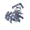
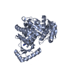
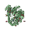
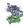
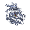
[English] 日本語
Yorodumi
- PDB-2vqd: Crystal Structure of Biotin Carboxylase from Pseudomonas aerugino... -

+
Open data


ID or keywords:

Loading...

-
Basic information

Entry
Database: PDB / ID: 2vqd
TitleCrystal Structure of Biotin Carboxylase from Pseudomonas aeruginosa complexed with AMPCP
ComponentsBIOTIN CARBOXYLASE
KeywordsLIGASE / NUCLEOTIDE-BINDING / FATTY ACID BIOSYNTHESIS / LIPID SYNTHESIS / ATP-GRASP DOMAIN / BIOTIN CARBOXYLASE / BIOTIN / BACTERIA / ATP-BINDING
Function / homology
Function and homology information


biotin carboxylase / biotin carboxylase activity / malonyl-CoA biosynthetic process / acetyl-CoA carboxylase activity / fatty acid biosynthetic process / ATP binding / metal ion binding
Similarity search - Function
Acetyl-CoA carboxylase, biotin carboxylase / Phosphoribosylglycinamide synthetase, ATP-grasp (A) domain / Biotin carboxylase-like, N-terminal domain / Biotin carboxylase, C-terminal / Biotin carboxylation domain / Biotin carboxylase, N-terminal domain / Biotin carboxylase C-terminal domain / Biotin carboxylation domain profile. / Biotin carboxylase C-terminal domain / Rossmann fold - #20 ...Acetyl-CoA carboxylase, biotin carboxylase / Phosphoribosylglycinamide synthetase, ATP-grasp (A) domain / Biotin carboxylase-like, N-terminal domain / Biotin carboxylase, C-terminal / Biotin carboxylation domain / Biotin carboxylase, N-terminal domain / Biotin carboxylase C-terminal domain / Biotin carboxylation domain profile. / Biotin carboxylase C-terminal domain / Rossmann fold - #20 / Carbamoyl-phosphate synthase subdomain signature 1. / Carbamoyl-phosphate synthetase large subunit-like, ATP-binding domain / Carbamoyl-phosphate synthase L chain, ATP binding domain / Rudiment single hybrid motif / ATP-grasp fold, A domain / ATP-grasp fold, B domain / ATP-grasp fold, subdomain 1 / Pre-ATP-grasp domain superfamily / ATP-grasp fold / ATP-grasp fold profile. / D-amino Acid Aminotransferase; Chain A, domain 1 / Dna Ligase; domain 1 / Carbamoyl-phosphate synthase subdomain signature 2. / Rossmann fold / 2-Layer Sandwich / 3-Layer(aba) Sandwich / Alpha Beta
Similarity search - Domain/homology
PHOSPHOMETHYLPHOSPHONIC ACID ADENOSYL ESTER / Biotin carboxylase
Similarity search - Component
Biological speciesPSEUDOMONAS AERUGINOSA (bacteria)
MethodX-RAY DIFFRACTION / MOLECULAR REPLACEMENT / Resolution: 2.41 Å
AuthorsMochalkin, I.
CitationJournal: Protein Sci. / Year: 2008
Title: Structural Evidence for Substrate-Induced Synergism and Half-Sites Reactivity in Biotin Carboxylase.
Authors: Mochalkin, I. / Miller, J.R. / Evdokimov, A. / Lightle, S. / Yan, C. / Stover, C.K. / Waldrop, G.L.
History
DepositionMar 13, 2008Deposition site: PDBE / Processing site: PDBE
Revision 1.0Sep 9, 2008Provider: repository / Type: Initial release
Revision 1.1Jul 13, 2011Group: Advisory / Version format compliance
Revision 1.2Jul 5, 2017Group: Data collection / Category: diffrn_source / Item: _diffrn_source.type
Revision 1.3Dec 13, 2023Group: Data collection / Database references ...Data collection / Database references / Derived calculations / Other / Refinement description
Category: chem_comp_atom / chem_comp_bond ...chem_comp_atom / chem_comp_bond / database_2 / pdbx_database_status / pdbx_initial_refinement_model / pdbx_struct_conn_angle / struct_conn
Item: _database_2.pdbx_DOI / _database_2.pdbx_database_accession ..._database_2.pdbx_DOI / _database_2.pdbx_database_accession / _pdbx_database_status.status_code_sf / _pdbx_struct_conn_angle.ptnr1_auth_comp_id / _pdbx_struct_conn_angle.ptnr1_auth_seq_id / _pdbx_struct_conn_angle.ptnr1_label_asym_id / _pdbx_struct_conn_angle.ptnr1_label_atom_id / _pdbx_struct_conn_angle.ptnr1_label_comp_id / _pdbx_struct_conn_angle.ptnr3_auth_comp_id / _pdbx_struct_conn_angle.ptnr3_auth_seq_id / _pdbx_struct_conn_angle.ptnr3_label_asym_id / _pdbx_struct_conn_angle.ptnr3_label_atom_id / _pdbx_struct_conn_angle.ptnr3_label_comp_id / _pdbx_struct_conn_angle.value / _struct_conn.pdbx_dist_value / _struct_conn.ptnr1_auth_comp_id / _struct_conn.ptnr1_auth_seq_id / _struct_conn.ptnr1_label_asym_id / _struct_conn.ptnr1_label_atom_id / _struct_conn.ptnr1_label_comp_id / _struct_conn.ptnr1_label_seq_id / _struct_conn.ptnr2_auth_comp_id / _struct_conn.ptnr2_auth_seq_id / _struct_conn.ptnr2_label_asym_id / _struct_conn.ptnr2_label_atom_id / _struct_conn.ptnr2_label_comp_id / _struct_conn.ptnr2_label_seq_id

-
Structure visualization

Structure viewerMolecule:
MolmilJmol/JSmol

Downloads & links

-
Assembly

Deposited unit
A: BIOTIN CARBOXYLASE
hetero molecules


Theoretical massNumber of molelcules
Total (without water)51,3254
Polymers50,7801
Non-polymers5463
Water4,179232
1


  • Idetical with deposited unit
  • defined by author&software
TypeNameSymmetry operationNumber
identity operation1_555x,y,z1
MethodPQS
Unit cell
Length a, b, c (Å)64.227, 126.786, 49.878
Angle α, β, γ (deg.)90.00, 90.00, 90.00
Int Tables number18
Space group name H-MP21212
Components on special symmetry positions
IDModelComponents
11A-2176-

HOH

-
Components

#1: Protein BIOTIN CARBOXYLASE / / ACETYL-COA CARBOXYLASE SUBUNIT A / ACC


Mass: 50779.715 Da / Num. of mol.: 1
Source method: isolated from a genetically manipulated source
Source: (gene. exp.) PSEUDOMONAS AERUGINOSA (bacteria) / Production host: ESCHERICHIA COLI (E. coli) / References: UniProt: P37798, biotin carboxylase
#2: Chemical ChemComp-AP2 / PHOSPHOMETHYLPHOSPHONIC ACID ADENOSYL ESTER


Mass: 425.228 Da / Num. of mol.: 1 / Source method: obtained synthetically / Formula: C11H17N5O9P2
#3: Chemical ChemComp-MG / MAGNESIUM ION


Mass: 24.305 Da / Num. of mol.: 1 / Source method: obtained synthetically / Formula: Mg
#4: Chemical ChemComp-SO4 / SULFATE ION / Sulfate


Mass: 96.063 Da / Num. of mol.: 1 / Source method: obtained synthetically / Formula: SO4
#5: Water ChemComp-HOH / water / Water


Mass: 18.015 Da / Num. of mol.: 232 / Source method: isolated from a natural source / Formula: H2O
Nonpolymer detailsPHOSPHOMETHYLPHOSPHONIC ACID ADENOSYL ESTER (AP2): NON-HYDROLYZABLE ADP ANALOG
Sequence detailsC-TERMINAL HIS-TAG PRESENT

-
Experimental details

-
Experiment

ExperimentMethod: X-RAY DIFFRACTION / Number of used crystals: 1

-
Sample preparation

CrystalDensity Matthews: 1.97 Å3/Da / Density % sol: 37.02 % / Description: NONE
Crystal growDetails: 0.1M HEPES (PH 7.0), 0.2M MGCL2, 15-20% PEG3350

-
Data collection

DiffractionMean temperature: 100 K
Diffraction sourceSource: ROTATING ANODE / Type: RIGAKU RUH2R / Wavelength: 1.54
DetectorDate: May 1, 2007
RadiationProtocol: SINGLE WAVELENGTH / Monochromatic (M) / Laue (L): M / Scattering type: x-ray
Radiation wavelengthWavelength: 1.54 Å / Relative weight: 1
ReflectionResolution: 2.4→20 Å / Num. obs: 15777 / % possible obs: 96 % / Observed criterion σ(I): 0 / Redundancy: 5.2 % / Rmerge(I) obs: 0.07 / Net I/σ(I): 22.9
Reflection shellResolution: 2.4→2.49 Å / Redundancy: 3.2 % / Rmerge(I) obs: 0.35 / Mean I/σ(I) obs: 2.86 / % possible all: 83.9

-
Processing

Software
NameVersionClassification
REFMAC5.2.0005refinement
HKL-2000data reduction
HKL-2000data scaling
MOLREPphasing
RefinementMethod to determine structure: MOLECULAR REPLACEMENT
Starting model: PDB ENTRY 1DV2
Resolution: 2.41→63.37 Å / Cor.coef. Fo:Fc: 0.953 / Cor.coef. Fo:Fc free: 0.916 / SU B: 18.358 / SU ML: 0.229 / TLS residual ADP flag: LIKELY RESIDUAL / Cross valid method: THROUGHOUT / ESU R: 1.859 / ESU R Free: 0.311 / Stereochemistry target values: MAXIMUM LIKELIHOOD / Details: HYDROGENS HAVE BEEN ADDED IN THE RIDING POSITIONS.
RfactorNum. reflection% reflectionSelection details
Rfree0.252 791 5 %RANDOM
Rwork0.178 ---
obs0.182 14953 95.7 %-
Solvent computationIon probe radii: 0.8 Å / Shrinkage radii: 0.8 Å / VDW probe radii: 1.4 Å / Solvent model: MASK
Displacement parametersBiso mean: 33.49 Å2
Baniso -1Baniso -2Baniso -3
1-1.51 Å20 Å20 Å2
2--0.21 Å20 Å2
3----1.73 Å2
Refinement stepCycle: LAST / Resolution: 2.41→63.37 Å
ProteinNucleic acidLigandSolventTotal
Num. atoms3412 0 33 232 3677
Refine LS restraints
Refine-IDTypeDev idealDev ideal targetNumber
X-RAY DIFFRACTIONr_bond_refined_d0.0090.0223514
X-RAY DIFFRACTIONr_bond_other_d0.0010.023247
X-RAY DIFFRACTIONr_angle_refined_deg1.2121.9814756
X-RAY DIFFRACTIONr_angle_other_deg0.75837541
X-RAY DIFFRACTIONr_dihedral_angle_1_deg5.8535448
X-RAY DIFFRACTIONr_dihedral_angle_2_deg36.8523.718156
X-RAY DIFFRACTIONr_dihedral_angle_3_deg15.60415600
X-RAY DIFFRACTIONr_dihedral_angle_4_deg16.9111529
X-RAY DIFFRACTIONr_chiral_restr0.0650.2529
X-RAY DIFFRACTIONr_gen_planes_refined0.0030.023943
X-RAY DIFFRACTIONr_gen_planes_other0.0010.02697
X-RAY DIFFRACTIONr_nbd_refined0.1880.2691
X-RAY DIFFRACTIONr_nbd_other0.1760.23225
X-RAY DIFFRACTIONr_nbtor_refined0.1650.21687
X-RAY DIFFRACTIONr_nbtor_other0.0820.22141
X-RAY DIFFRACTIONr_xyhbond_nbd_refined0.1490.2176
X-RAY DIFFRACTIONr_xyhbond_nbd_other
X-RAY DIFFRACTIONr_metal_ion_refined
X-RAY DIFFRACTIONr_metal_ion_other
X-RAY DIFFRACTIONr_symmetry_vdw_refined0.1470.217
X-RAY DIFFRACTIONr_symmetry_vdw_other0.1820.284
X-RAY DIFFRACTIONr_symmetry_hbond_refined0.1720.219
X-RAY DIFFRACTIONr_symmetry_hbond_other
X-RAY DIFFRACTIONr_symmetry_metal_ion_refined
X-RAY DIFFRACTIONr_symmetry_metal_ion_other
X-RAY DIFFRACTIONr_mcbond_it0.5431.52883
X-RAY DIFFRACTIONr_mcbond_other
X-RAY DIFFRACTIONr_mcangle_it0.61923558
X-RAY DIFFRACTIONr_mcangle_other
X-RAY DIFFRACTIONr_scbond_it1.02131469
X-RAY DIFFRACTIONr_scbond_other
X-RAY DIFFRACTIONr_scangle_it1.5034.51198
X-RAY DIFFRACTIONr_scangle_other
X-RAY DIFFRACTIONr_long_range_B_refined
X-RAY DIFFRACTIONr_long_range_B_other
X-RAY DIFFRACTIONr_rigid_bond_restr
X-RAY DIFFRACTIONr_sphericity_free
X-RAY DIFFRACTIONr_sphericity_bonded
LS refinement shellResolution: 2.41→2.47 Å / Total num. of bins used: 20 /
RfactorNum. reflection
Rfree0.321 44
Rwork0.224 884
Refinement TLS params.

Method: refined / Refine-ID: X-RAY DIFFRACTION

IDL112)L122)L132)L222)L232)L332)S11 (Å °)S12 (Å °)S13 (Å °)S21 (Å °)S22 (Å °)S23 (Å °)S31 (Å °)S32 (Å °)S33 (Å °)T112)T122)T132)T222)T232)T332)Origin x (Å)Origin y (Å)Origin z (Å)
10.59960.0165-0.14971.9352-0.39720.35160.03930.01070.08270.211-0.042-0.114-0.0783-0.00120.0027-0.0552-0.0093-0.0309-0.0555-0.017-0.09936.121920.344913.0923
219.7093-0.360517.168730.05040.822814.99860.6192-0.51990.7931-0.1356-0.80251.4562-0.24690.40230.18330.0057-0.0160.00670.0004-0.00960.00310.335831.08489.8703
Refinement TLS group
IDRefine-IDRefine TLS-IDAuth asym-IDAuth seq-ID
1X-RAY DIFFRACTION1A1 - 447
2X-RAY DIFFRACTION2A1448

+
About Yorodumi

-
News

-
Feb 9, 2022. New format data for meta-information of EMDB entries

New format data for meta-information of EMDB entries

  • Version 3 of the EMDB header file is now the official format.
  • The previous official version 1.9 will be removed from the archive.

Related info.:EMDB header

External links:wwPDB to switch to version 3 of the EMDB data model

-
Aug 12, 2020. Covid-19 info

Covid-19 info

URL: https://pdbj.org/emnavi/covid19.php

New page: Covid-19 featured information page in EM Navigator.

Related info.:Covid-19 info / Mar 5, 2020. Novel coronavirus structure data

+
Mar 5, 2020. Novel coronavirus structure data

Novel coronavirus structure data

Related info.:Yorodumi Speices / Aug 12, 2020. Covid-19 info

External links:COVID-19 featured content - PDBj / Molecule of the Month (242):Coronavirus Proteases

+
Jan 31, 2019. EMDB accession codes are about to change! (news from PDBe EMDB page)

EMDB accession codes are about to change! (news from PDBe EMDB page)

  • The allocation of 4 digits for EMDB accession codes will soon come to an end. Whilst these codes will remain in use, new EMDB accession codes will include an additional digit and will expand incrementally as the available range of codes is exhausted. The current 4-digit format prefixed with “EMD-” (i.e. EMD-XXXX) will advance to a 5-digit format (i.e. EMD-XXXXX), and so on. It is currently estimated that the 4-digit codes will be depleted around Spring 2019, at which point the 5-digit format will come into force.
  • The EM Navigator/Yorodumi systems omit the EMD- prefix.

Related info.:Q: What is EMD? / ID/Accession-code notation in Yorodumi/EM Navigator

External links:EMDB Accession Codes are Changing Soon! / Contact to PDBj

+
Jul 12, 2017. Major update of PDB

Major update of PDB

  • wwPDB released updated PDB data conforming to the new PDBx/mmCIF dictionary.
  • This is a major update changing the version number from 4 to 5, and with Remediation, in which all the entries are updated.
  • In this update, many items about electron microscopy experimental information are reorganized (e.g. em_software).
  • Now, EM Navigator and Yorodumi are based on the updated data.

External links:wwPDB Remediation / Enriched Model Files Conforming to OneDep Data Standards Now Available in the PDB FTP Archive

-
Yorodumi

Thousand views of thousand structures

  • Yorodumi is a browser for structure data from EMDB, PDB, SASBDB, etc.
  • This page is also the successor to EM Navigator detail page, and also detail information page/front-end page for Omokage search.
  • The word "yorodu" (or yorozu) is an old Japanese word meaning "ten thousand". "mi" (miru) is to see.

Related info.:EMDB / PDB / SASBDB / Comparison of 3 databanks / Yorodumi Search / Aug 31, 2016. New EM Navigator & Yorodumi / Yorodumi Papers / Jmol/JSmol / Function and homology information / Changes in new EM Navigator and Yorodumi

Read more