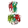
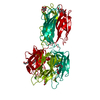
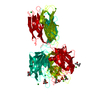
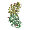
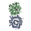
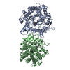
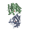
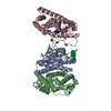
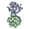
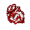
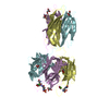
[English] 日本語
Yorodumi
- PDB-2vme: Structure of the wild-type discoidin II from Dictyostelium discoideum -

+
Open data


ID or keywords:

Loading...

-
Basic information

Entry
Database: PDB / ID: 2vme
TitleStructure of the wild-type discoidin II from Dictyostelium discoideum
ComponentsDISCOIDIN-2Discoidin domain
KeywordsCELL ADHESION / LECTIN / AGGREGATION / DDR
Function / homology
Function and homology information


adenylate cyclase-activating G protein-coupled cAMP receptor signaling pathway / spore wall / N-acetylgalactosamine binding / protein complex involved in cell adhesion / galactoside binding / oligosaccharide binding / polysaccharide binding / phagocytic vesicle / cytoskeleton organization / extracellular matrix ...adenylate cyclase-activating G protein-coupled cAMP receptor signaling pathway / spore wall / N-acetylgalactosamine binding / protein complex involved in cell adhesion / galactoside binding / oligosaccharide binding / polysaccharide binding / phagocytic vesicle / cytoskeleton organization / extracellular matrix / response to bacterium / cell-cell adhesion / carbohydrate binding / vesicle / cell adhesion / metal ion binding / cytosol / cytoplasm
Similarity search - Function
Immunoglobulin-like - #2080 / H-type lectin domain / H-type lectin domain superfamily / H-type lectin domain / Coagulation factors 5/8 type C domain (FA58C) signature 1. / Coagulation factor 5/8 C-terminal domain, discoidin domain / Coagulation factors 5/8 type C domain (FA58C) profile. / F5/8 type C domain / Coagulation factor 5/8 C-terminal domain / Galactose-binding domain-like ...Immunoglobulin-like - #2080 / H-type lectin domain / H-type lectin domain superfamily / H-type lectin domain / Coagulation factors 5/8 type C domain (FA58C) signature 1. / Coagulation factor 5/8 C-terminal domain, discoidin domain / Coagulation factors 5/8 type C domain (FA58C) profile. / F5/8 type C domain / Coagulation factor 5/8 C-terminal domain / Galactose-binding domain-like / Galactose-binding-like domain superfamily / Jelly Rolls / Immunoglobulin-like / Sandwich / Mainly Beta
Similarity search - Domain/homology
Biological speciesDICTYOSTELIUM DISCOIDEUM (eukaryote)
MethodX-RAY DIFFRACTION / SYNCHROTRON / MOLECULAR REPLACEMENT / Resolution: 2.45 Å
AuthorsAragao, K.S. / Satre, M. / Imberty, A. / Varrot, A.
CitationJournal: Proteins / Year: 2008
Title: Structure Determination of Discoidin II from Dictyostelium Discoideum and Carbohydrate Binding Properties of the Lectin Domain.
Authors: Aragao, K.S. / Satre, M. / Imberty, A. / Varrot, A.
History
DepositionJan 25, 2008Deposition site: PDBE / Processing site: PDBE
Revision 1.0Jul 1, 2008Provider: repository / Type: Initial release
Revision 1.1Jun 2, 2011Group: Version format compliance
Revision 1.2Jul 13, 2011Group: Version format compliance
Revision 1.3Dec 13, 2023Group: Data collection / Database references ...Data collection / Database references / Derived calculations / Other / Refinement description
Category: chem_comp_atom / chem_comp_bond ...chem_comp_atom / chem_comp_bond / database_2 / pdbx_database_status / pdbx_initial_refinement_model / pdbx_struct_conn_angle / struct_conn / struct_site
Item: _database_2.pdbx_DOI / _database_2.pdbx_database_accession ..._database_2.pdbx_DOI / _database_2.pdbx_database_accession / _pdbx_database_status.status_code_sf / _pdbx_struct_conn_angle.ptnr1_auth_asym_id / _pdbx_struct_conn_angle.ptnr1_auth_comp_id / _pdbx_struct_conn_angle.ptnr1_auth_seq_id / _pdbx_struct_conn_angle.ptnr1_label_asym_id / _pdbx_struct_conn_angle.ptnr1_label_atom_id / _pdbx_struct_conn_angle.ptnr1_label_comp_id / _pdbx_struct_conn_angle.ptnr1_label_seq_id / _pdbx_struct_conn_angle.ptnr3_auth_asym_id / _pdbx_struct_conn_angle.ptnr3_auth_comp_id / _pdbx_struct_conn_angle.ptnr3_auth_seq_id / _pdbx_struct_conn_angle.ptnr3_label_asym_id / _pdbx_struct_conn_angle.ptnr3_label_atom_id / _pdbx_struct_conn_angle.ptnr3_label_comp_id / _pdbx_struct_conn_angle.ptnr3_label_seq_id / _pdbx_struct_conn_angle.value / _struct_conn.conn_type_id / _struct_conn.id / _struct_conn.pdbx_dist_value / _struct_conn.pdbx_leaving_atom_flag / _struct_conn.ptnr1_auth_asym_id / _struct_conn.ptnr1_auth_comp_id / _struct_conn.ptnr1_auth_seq_id / _struct_conn.ptnr1_label_asym_id / _struct_conn.ptnr1_label_atom_id / _struct_conn.ptnr1_label_comp_id / _struct_conn.ptnr1_label_seq_id / _struct_conn.ptnr2_auth_asym_id / _struct_conn.ptnr2_auth_comp_id / _struct_conn.ptnr2_auth_seq_id / _struct_conn.ptnr2_label_asym_id / _struct_conn.ptnr2_label_atom_id / _struct_conn.ptnr2_label_comp_id / _struct_conn.ptnr2_label_seq_id / _struct_site.pdbx_auth_asym_id / _struct_site.pdbx_auth_comp_id / _struct_site.pdbx_auth_seq_id
Remark 650 HELIX DETERMINATION METHOD: AUTHOR PROVIDED.
Remark 700 SHEET DETERMINATION METHOD: AUTHOR PROVIDED.

-
Structure visualization

Structure viewerMolecule:
MolmilJmol/JSmol

Downloads & links

-
Assembly

Deposited unit
A: DISCOIDIN-2
B: DISCOIDIN-2
C: DISCOIDIN-2
D: DISCOIDIN-2
E: DISCOIDIN-2
F: DISCOIDIN-2
hetero molecules


Theoretical massNumber of molelcules
Total (without water)174,13232
Polymers172,1146
Non-polymers2,01826
Water9,782543
1
A: DISCOIDIN-2
B: DISCOIDIN-2
C: DISCOIDIN-2
hetero molecules


Theoretical massNumber of molelcules
Total (without water)87,11716
Polymers86,0573
Non-polymers1,06013
Water543
TypeNameSymmetry operationNumber
identity operation1_555x,y,z1
Buried area14210 Å2
ΔGint-64.4 kcal/mol
Surface area28410 Å2
MethodPISA
2
D: DISCOIDIN-2
E: DISCOIDIN-2
F: DISCOIDIN-2
hetero molecules


Theoretical massNumber of molelcules
Total (without water)87,01516
Polymers86,0573
Non-polymers95813
Water543
TypeNameSymmetry operationNumber
identity operation1_555x,y,z1
Buried area13990 Å2
ΔGint-71.1 kcal/mol
Surface area28530 Å2
MethodPISA
Unit cell
Length a, b, c (Å)287.885, 56.281, 123.674
Angle α, β, γ (deg.)90.00, 109.10, 90.00
Int Tables number5
Space group name H-MC121

-
Components

-
Protein , 1 types, 6 molecules ABCDEF

#1: Protein
DISCOIDIN-2 / Discoidin domain / DISCOIDIN II


Mass: 28685.703 Da / Num. of mol.: 6 / Source method: isolated from a natural source / Details: HIS84 IS PHOSPHORYLATED. / Source: (natural) DICTYOSTELIUM DISCOIDEUM (eukaryote) / Strain: AX2 / References: UniProt: P42530

-
Non-polymers , 6 types, 569 molecules

#2: Chemical
ChemComp-CA / CALCIUM ION


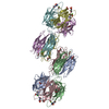
Mass: 40.078 Da / Num. of mol.: 6 / Source method: obtained synthetically / Formula: Ca
#3: Chemical ChemComp-ZN / ZINC ION


Mass: 65.409 Da / Num. of mol.: 2 / Source method: obtained synthetically / Formula: Zn
#4: Chemical ChemComp-CL / CHLORIDE ION / Chloride


Mass: 35.453 Da / Num. of mol.: 2 / Source method: obtained synthetically / Formula: Cl
#5: Chemical
ChemComp-GOL / GLYCEROL / GLYCERIN / PROPANE-1,2,3-TRIOL / Glycerol


Mass: 92.094 Da / Num. of mol.: 15 / Source method: obtained synthetically / Formula: C3H8O3
#6: Chemical ChemComp-PG4 / TETRAETHYLENE GLYCOL / Polyethylene glycol


Mass: 194.226 Da / Num. of mol.: 1 / Source method: obtained synthetically / Formula: C8H18O5 / Comment: precipitant*YM
#7: Water ChemComp-HOH / water / Water


Mass: 18.015 Da / Num. of mol.: 543 / Source method: isolated from a natural source / Formula: H2O

-
Details

Nonpolymer detailsGLYCEROL (GOL): FROM CRYOPROTECTANT

-
Experimental details

-
Experiment

ExperimentMethod: X-RAY DIFFRACTION / Number of used crystals: 1

-
Sample preparation

CrystalDensity Matthews: 2.73 Å3/Da / Density % sol: 54.95 % / Description: NONE
Crystal growpH: 8.5 / Details: 20% PEG 4K, 10 MM CDCL2, 100 MM HEPES PH 7.5

-
Data collection

DiffractionMean temperature: 100 K
Diffraction sourceSource: SYNCHROTRON / Site: ESRF / Beamline: ID23-1 / Wavelength: 0.976
DetectorType: ADSC CCD / Detector: CCD / Date: Feb 10, 2006
RadiationProtocol: SINGLE WAVELENGTH / Monochromatic (M) / Laue (L): M / Scattering type: x-ray
Radiation wavelengthWavelength: 0.976 Å / Relative weight: 1
ReflectionResolution: 2.45→26.82 Å / Num. obs: 66709 / % possible obs: 96.8 % / Observed criterion σ(I): 2 / Redundancy: 3.2 % / Rmerge(I) obs: 0.09 / Net I/σ(I): 5.8
Reflection shellResolution: 2.45→2.58 Å / Redundancy: 3.1 % / Rmerge(I) obs: 0.37 / Mean I/σ(I) obs: 1.9 / % possible all: 98.3

-
Processing

Software
NameVersionClassification
REFMAC5.2.0019refinement
MOSFLMdata reduction
SCALAdata scaling
PHASERphasing
RefinementMethod to determine structure: MOLECULAR REPLACEMENT
Starting model: PDB ENTRY 2VM9
Resolution: 2.45→26.8 Å / Cor.coef. Fo:Fc: 0.943 / Cor.coef. Fo:Fc free: 0.903 / SU B: 8.226 / SU ML: 0.185 / Cross valid method: THROUGHOUT / ESU R: 0.467 / ESU R Free: 0.271 / Stereochemistry target values: MAXIMUM LIKELIHOOD
Details: HYDROGENS HAVE BEEN ADDED IN THE RIDING POSITIONS. DISORDERED SIDE CHAINS WERE MODELED STEREOCHEMICALLY
RfactorNum. reflection% reflectionSelection details
Rfree0.238 3380 5.1 %RANDOM
Rwork0.181 ---
obs0.184 63327 95.9 %-
Solvent computationIon probe radii: 0.8 Å / Shrinkage radii: 0.8 Å / VDW probe radii: 1.2 Å / Solvent model: MASK
Displacement parametersBiso mean: 26.78 Å2
Baniso -1Baniso -2Baniso -3
1--1.5 Å20 Å2-0.74 Å2
2--0.99 Å20 Å2
3---0.03 Å2
Refinement stepCycle: LAST / Resolution: 2.45→26.8 Å
ProteinNucleic acidLigandSolventTotal
Num. atoms12084 0 113 543 12740
Refine LS restraints
Refine-IDTypeDev idealDev ideal targetNumber
X-RAY DIFFRACTIONr_bond_refined_d0.0140.02112504
X-RAY DIFFRACTIONr_bond_other_d
X-RAY DIFFRACTIONr_angle_refined_deg1.5561.93116988
X-RAY DIFFRACTIONr_angle_other_deg
X-RAY DIFFRACTIONr_dihedral_angle_1_deg7.46451538
X-RAY DIFFRACTIONr_dihedral_angle_2_deg39.11824.577603
X-RAY DIFFRACTIONr_dihedral_angle_3_deg15.283151965
X-RAY DIFFRACTIONr_dihedral_angle_4_deg20.2181566
X-RAY DIFFRACTIONr_chiral_restr0.10.21863
X-RAY DIFFRACTIONr_gen_planes_refined0.0050.029568
X-RAY DIFFRACTIONr_gen_planes_other
X-RAY DIFFRACTIONr_nbd_refined0.2120.25682
X-RAY DIFFRACTIONr_nbd_other
X-RAY DIFFRACTIONr_nbtor_refined0.3110.28441
X-RAY DIFFRACTIONr_nbtor_other
X-RAY DIFFRACTIONr_xyhbond_nbd_refined0.1490.2869
X-RAY DIFFRACTIONr_xyhbond_nbd_other
X-RAY DIFFRACTIONr_metal_ion_refined
X-RAY DIFFRACTIONr_metal_ion_other
X-RAY DIFFRACTIONr_symmetry_vdw_refined0.2260.232
X-RAY DIFFRACTIONr_symmetry_vdw_other
X-RAY DIFFRACTIONr_symmetry_hbond_refined0.0460.23
X-RAY DIFFRACTIONr_symmetry_hbond_other
X-RAY DIFFRACTIONr_symmetry_metal_ion_refined
X-RAY DIFFRACTIONr_symmetry_metal_ion_other
X-RAY DIFFRACTIONr_mcbond_it0.6821.57792
X-RAY DIFFRACTIONr_mcbond_other
X-RAY DIFFRACTIONr_mcangle_it1.185212406
X-RAY DIFFRACTIONr_mcangle_other
X-RAY DIFFRACTIONr_scbond_it1.85135351
X-RAY DIFFRACTIONr_scbond_other
X-RAY DIFFRACTIONr_scangle_it2.6894.54565
X-RAY DIFFRACTIONr_scangle_other
X-RAY DIFFRACTIONr_long_range_B_refined
X-RAY DIFFRACTIONr_long_range_B_other
X-RAY DIFFRACTIONr_rigid_bond_restr
X-RAY DIFFRACTIONr_sphericity_free
X-RAY DIFFRACTIONr_sphericity_bonded
LS refinement shellResolution: 2.45→2.51 Å / Total num. of bins used: 20 /
RfactorNum. reflection
Rfree0.329 267
Rwork0.249 4666

+
About Yorodumi

-
News

-
Feb 9, 2022. New format data for meta-information of EMDB entries

New format data for meta-information of EMDB entries

  • Version 3 of the EMDB header file is now the official format.
  • The previous official version 1.9 will be removed from the archive.

Related info.:EMDB header

External links:wwPDB to switch to version 3 of the EMDB data model

-
Aug 12, 2020. Covid-19 info

Covid-19 info

URL: https://pdbj.org/emnavi/covid19.php

New page: Covid-19 featured information page in EM Navigator.

Related info.:Covid-19 info / Mar 5, 2020. Novel coronavirus structure data

+
Mar 5, 2020. Novel coronavirus structure data

Novel coronavirus structure data

Related info.:Yorodumi Speices / Aug 12, 2020. Covid-19 info

External links:COVID-19 featured content - PDBj / Molecule of the Month (242):Coronavirus Proteases

+
Jan 31, 2019. EMDB accession codes are about to change! (news from PDBe EMDB page)

EMDB accession codes are about to change! (news from PDBe EMDB page)

  • The allocation of 4 digits for EMDB accession codes will soon come to an end. Whilst these codes will remain in use, new EMDB accession codes will include an additional digit and will expand incrementally as the available range of codes is exhausted. The current 4-digit format prefixed with “EMD-” (i.e. EMD-XXXX) will advance to a 5-digit format (i.e. EMD-XXXXX), and so on. It is currently estimated that the 4-digit codes will be depleted around Spring 2019, at which point the 5-digit format will come into force.
  • The EM Navigator/Yorodumi systems omit the EMD- prefix.

Related info.:Q: What is EMD? / ID/Accession-code notation in Yorodumi/EM Navigator

External links:EMDB Accession Codes are Changing Soon! / Contact to PDBj

+
Jul 12, 2017. Major update of PDB

Major update of PDB

  • wwPDB released updated PDB data conforming to the new PDBx/mmCIF dictionary.
  • This is a major update changing the version number from 4 to 5, and with Remediation, in which all the entries are updated.
  • In this update, many items about electron microscopy experimental information are reorganized (e.g. em_software).
  • Now, EM Navigator and Yorodumi are based on the updated data.

External links:wwPDB Remediation / Enriched Model Files Conforming to OneDep Data Standards Now Available in the PDB FTP Archive

-
Yorodumi

Thousand views of thousand structures

  • Yorodumi is a browser for structure data from EMDB, PDB, SASBDB, etc.
  • This page is also the successor to EM Navigator detail page, and also detail information page/front-end page for Omokage search.
  • The word "yorodu" (or yorozu) is an old Japanese word meaning "ten thousand". "mi" (miru) is to see.

Related info.:EMDB / PDB / SASBDB / Comparison of 3 databanks / Yorodumi Search / Aug 31, 2016. New EM Navigator & Yorodumi / Yorodumi Papers / Jmol/JSmol / Function and homology information / Changes in new EM Navigator and Yorodumi

Read more