 Movie
Movie Controller


+ Open data

- Basic information
| Entry | Database: PDB / ID: 2vfj | ||||||
|---|---|---|---|---|---|---|---|
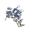
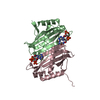
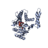
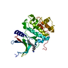
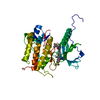
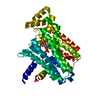
| Title | Structure of the A20 Ovarian Tumour (OTU) domain | ||||||
 Components Components | TUMOR NECROSIS FACTOR | ||||||
 Keywords Keywords | HYDROLASE / PHOSPHORYLATION / CYSTEINE PROTEASE / METAL-BINDING / OVARIAN TUMOUR / THIOL PROTEASE / DNA-BINDING / POLYMORPHISM / LYS63-LINKED / CYTOPLASM / UBIQUITIN / ZINC-FINGER / DEUBIQUITINATING ENZYME / CYTOKINE SIGNALLING / UBL CONJUGATION PATHWAY / OTU / ZINC / NF-KB / NUCLEUS / PROTEASE / APOPTOSIS | ||||||
| Function / homology |  Function and homology information Function and homology informationregulation of vascular wound healing / negative regulation of toll-like receptor 5 signaling pathway / negative regulation of nucleotide-binding oligomerization domain containing 1 signaling pathway / establishment of protein localization to vacuole / negative regulation of osteoclast proliferation / tolerance induction to lipopolysaccharide / negative regulation of CD40 signaling pathway / negative regulation of toll-like receptor 3 signaling pathway / negative regulation of B cell activation / negative regulation of nucleotide-binding oligomerization domain containing 2 signaling pathway ...regulation of vascular wound healing / negative regulation of toll-like receptor 5 signaling pathway / negative regulation of nucleotide-binding oligomerization domain containing 1 signaling pathway / establishment of protein localization to vacuole / negative regulation of osteoclast proliferation / tolerance induction to lipopolysaccharide / negative regulation of CD40 signaling pathway / negative regulation of toll-like receptor 3 signaling pathway / negative regulation of B cell activation / negative regulation of nucleotide-binding oligomerization domain containing 2 signaling pathway / negative regulation of toll-like receptor 2 signaling pathway / protein K11-linked deubiquitination / nucleotide-binding domain, leucine rich repeat containing receptor signaling pathway / negative regulation of chronic inflammatory response / negative regulation of toll-like receptor 4 signaling pathway / regulation of germinal center formation / protein K48-linked deubiquitination / B-1 B cell homeostasis / regulation of defense response to virus by host / regulation of tumor necrosis factor-mediated signaling pathway / Transferases; Acyltransferases; Aminoacyltransferases / protein K63-linked deubiquitination / positive regulation of hepatocyte proliferation / negative regulation of bone resorption / TNFR1-induced proapoptotic signaling / negative regulation of interleukin-1 beta production / negative regulation of interleukin-2 production / K63-linked polyubiquitin modification-dependent protein binding / K63-linked deubiquitinase activity / negative regulation of NF-kappaB transcription factor activity / protein deubiquitination / negative regulation of interleukin-6 production / response to muramyl dipeptide / negative regulation of tumor necrosis factor production / positive regulation of Wnt signaling pathway / protein K48-linked ubiquitination / negative regulation of endothelial cell apoptotic process / negative regulation of canonical NF-kappaB signal transduction / negative regulation of extrinsic apoptotic signaling pathway via death domain receptors / negative regulation of protein ubiquitination / cytoskeleton organization / negative regulation of innate immune response / TNFR1-induced NF-kappa-B signaling pathway / ubiquitin binding / Negative regulators of DDX58/IFIH1 signaling / negative regulation of smooth muscle cell proliferation / Regulation of TNFR1 signaling / NOD1/2 Signaling Pathway / response to molecule of bacterial origin / kinase binding / negative regulation of inflammatory response / cellular response to hydrogen peroxide / positive regulation of protein catabolic process / ubiquitin-protein transferase activity / : / Ovarian tumor domain proteases / cell migration / protease binding / cellular response to lipopolysaccharide / ubiquitinyl hydrolase 1 / cysteine-type deubiquitinase activity / lysosome / inflammatory response / apoptotic process / proteolysis / DNA binding / extracellular exosome / zinc ion binding / identical protein binding / nucleus / cytosol / cytoplasmSimilarity search - Function | ||||||
| Biological species |  HOMO SAPIENS (human) HOMO SAPIENS (human) | ||||||
| Method | X-RAY DIFFRACTION / SYNCHROTRON / MAD / Resolution: 3.2 Å | ||||||
 Authors Authors | Komander, D. / Barford, D. | ||||||
 Citation Citation | Journal: Biochem.J. / Year: 2008 Title: Structure of the A20 Otu Domain and Mechanistic Insights Into Deubiquitination Authors: Komander, D. / Barford, D. | ||||||
| History |
|
- Structure visualization
| Structure viewer | Molecule: MolmilJmol/JSmol |
|---|
- Downloads & links
-Download
| PDBx/mmCIF format | 2vfj.cif.gz | 526.5 KB | Display | PDBx/mmCIF format |
|---|---|---|---|---|
| PDB format | pdb2vfj.ent.gz | 438.5 KB | Display | PDB format |
| PDBx/mmJSON format | 2vfj.json.gz | Tree view | PDBx/mmJSON format | |
| Others |  Other downloads Other downloads |
-Validation report
| Arichive directory | https://data.pdbj.org/pub/pdb/validation_reports/vf/2vfjftp://data.pdbj.org/pub/pdb/validation_reports/vf/2vfj | HTTPS FTP |
|---|
-Related structure data
| Similar structure data |
|---|


-Links
 PDBj
PDBj


- Assembly
| Deposited unit | 
| ||||||||
|---|---|---|---|---|---|---|---|---|---|
| 1 | 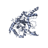
| ||||||||
| 2 | 
| ||||||||
| 3 | 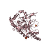
| ||||||||
| 4 | 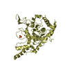
| ||||||||
| Unit cell |
|
-Components
| #1: Protein | / ALPHA-INDUCED PROTEIN 3 / DNA-BINDING PROTEIN A20 / ZINC FINGER PROTEIN A20 Mass: 43103.480 Da / Num. of mol.: 4 / Fragment: OVARIAN TUMOUR (OTU) DOMAIN, RESIDUES 1-366 Source method: isolated from a genetically manipulated source Details: RESIDUES 1-366 / Source: (gene. exp.) HOMO SAPIENS (human) / Plasmid: PGEX6P1 / Production host:  ESCHERICHIA COLI (E. coli) / Strain (production host): BL21 DE3 / References: UniProt: P21580, ubiquitinyl hydrolase 1 ESCHERICHIA COLI (E. coli) / Strain (production host): BL21 DE3 / References: UniProt: P21580, ubiquitinyl hydrolase 1#2: Chemical | ChemComp-SO4 / Sulfate  Mass: 96.063 Da / Num. of mol.: 4 / Source method: obtained synthetically / Formula: SO4 Mass: 96.063 Da / Num. of mol.: 4 / Source method: obtained synthetically / Formula: SO4#3: Chemical | ChemComp-MG / |   Mass: 24.305 Da / Num. of mol.: 1 / Source method: obtained synthetically / Formula: Mg Mass: 24.305 Da / Num. of mol.: 1 / Source method: obtained synthetically / Formula: MgSequence details | RESIDUES 1-366 OF A20 SEVERAL RESIDUES WERE MODELLED AS ALA DUE TO DISORDER | |
|---|
-Experimental details
-Experiment
| Experiment | Method: X-RAY DIFFRACTION / Number of used crystals: 1 |
|---|
- Sample preparation
| Crystal | Density Matthews: 3.4 Å3/Da / Density % sol: 63 % / Description: 3 WAVELENGTH MAD EXPERIMENT |
|---|---|
| Crystal grow | Details: 1.3-1.6 M MAGNESIUM SULPHATE, 0.1 M MES [PH 6.5-6.9] |
-Data collection
| Diffraction | Mean temperature: 100 K |
|---|---|
| Diffraction source | Source: SYNCHROTRON / Site: ESRF  / Beamline: ID14-2 / Wavelength: 0.934 / Beamline: ID14-2 / Wavelength: 0.934 |
| Detector | Type: ADSC CCD / Detector: CCD / Date: Oct 10, 2005 |
| Radiation | Protocol: SINGLE WAVELENGTH / Monochromatic (M) / Laue (L): M / Scattering type: x-ray |
| Radiation wavelength | Wavelength: 0.934 Å / Relative weight: 1 |
| Reflection | Resolution: 3.2→50 Å / Num. obs: 36226 / % possible obs: 96 % / Observed criterion σ(I): 2 / Redundancy: 4.2 % / Rmerge(I) obs: 0.08 / Net I/σ(I): 15.9 |
| Reflection shell | Resolution: 3.2→3.37 Å / Redundancy: 4.3 % / Rmerge(I) obs: 0.56 / Mean I/σ(I) obs: 2.6 / % possible all: 96 |
- Processing
| Software |
| |||||||||||||||||||||||||||||||||||||||||||||||||||||||||||||||||||||||||||||||||||||||||||||||||||||||||||||||||||||||||||||
|---|---|---|---|---|---|---|---|---|---|---|---|---|---|---|---|---|---|---|---|---|---|---|---|---|---|---|---|---|---|---|---|---|---|---|---|---|---|---|---|---|---|---|---|---|---|---|---|---|---|---|---|---|---|---|---|---|---|---|---|---|---|---|---|---|---|---|---|---|---|---|---|---|---|---|---|---|---|---|---|---|---|---|---|---|---|---|---|---|---|---|---|---|---|---|---|---|---|---|---|---|---|---|---|---|---|---|---|---|---|---|---|---|---|---|---|---|---|---|---|---|---|---|---|---|---|---|
| Refinement | Method to determine structure: MAD Starting model: NONE Resolution: 3.2→50.286 Å / σ(F): 1.4 / Stereochemistry target values: ML Details: THE ANISO RECORDS CORRESPOND TO TLS FACTORS INCORPORATED BY PHENIX AND NOT ANISOTROPIC REFINEMENT OF INDIVIDUAL ATOMS.
| |||||||||||||||||||||||||||||||||||||||||||||||||||||||||||||||||||||||||||||||||||||||||||||||||||||||||||||||||||||||||||||
| Refinement step | Cycle: LAST / Resolution: 3.2→50.286 Å
| |||||||||||||||||||||||||||||||||||||||||||||||||||||||||||||||||||||||||||||||||||||||||||||||||||||||||||||||||||||||||||||
| Refine LS restraints |
| |||||||||||||||||||||||||||||||||||||||||||||||||||||||||||||||||||||||||||||||||||||||||||||||||||||||||||||||||||||||||||||
| Refinement TLS params. | Method: refined / Refine-ID: X-RAY DIFFRACTION
| |||||||||||||||||||||||||||||||||||||||||||||||||||||||||||||||||||||||||||||||||||||||||||||||||||||||||||||||||||||||||||||
| Refinement TLS group |
|
