 Movie
Movie Controller


+ Open data

- Basic information
| Entry | Database: PDB / ID: 2v8n | ||||||
|---|---|---|---|---|---|---|---|
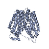
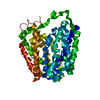
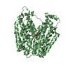
| Title | Wild-type Structure of Lactose Permease | ||||||
 Components Components | LACTOSE PERMEASE | ||||||
 Keywords Keywords | TRANSPORT PROTEIN / TRANSMEMBRANE / INNER MEMBRANE / SUGAR TRANSPORT / SYMPORT / MEMBRANE / TRANSPORT / FORMYLATION | ||||||
| Function / homology |  Function and homology information Function and homology informationlactose:proton symporter activity / lactose transport / carbohydrate:proton symporter activity / lactose binding / organic substance transport / carbohydrate transport / membrane / plasma membraneSimilarity search - Function | ||||||
| Biological species |  ESCHERICHIA COLI (E. coli) ESCHERICHIA COLI (E. coli) | ||||||
| Method | X-RAY DIFFRACTION / SYNCHROTRON / MOLECULAR REPLACEMENT / Resolution: 3.6 Å | ||||||
 Authors Authors | Guan, L. / Mirza, O. / Verner, G. / Iwata, S. / Kaback, H.R. | ||||||
 Citation Citation | Journal: Proc.Natl.Acad.Sci.USA / Year: 2007 Title: Structural Determination of Wild-Type Lactose Permease. Authors: Guan, L. / Mirza, O. / Verner, G. / Iwata, S. / Kaback, H.R. | ||||||
| History |
|
- Structure visualization
| Structure viewer | Molecule: MolmilJmol/JSmol |
|---|
- Downloads & links
-Download
| PDBx/mmCIF format | 2v8n.cif.gz | 170 KB | Display | PDBx/mmCIF format |
|---|---|---|---|---|
| PDB format | pdb2v8n.ent.gz | 136.7 KB | Display | PDB format |
| PDBx/mmJSON format | 2v8n.json.gz | Tree view | PDBx/mmJSON format | |
| Others |  Other downloads Other downloads |
-Validation report
| Arichive directory | https://data.pdbj.org/pub/pdb/validation_reports/v8/2v8nftp://data.pdbj.org/pub/pdb/validation_reports/v8/2v8n | HTTPS FTP |
|---|
-Related structure data
| Related structure data |  1pv7S S: Starting model for refinement |
|---|---|
| Similar structure data |


-Links
 PDBj
PDBj


- Assembly
| Deposited unit | 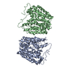
| ||||||||
|---|---|---|---|---|---|---|---|---|---|
| 1 | 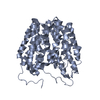
| ||||||||
| 2 | 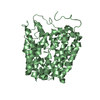
| ||||||||
| Unit cell |
| ||||||||
| Noncrystallographic symmetry (NCS) | NCS oper: (Code: given Matrix: (-0.004175, 1, -5.2E-5), Vector : |
-Components
| #1: Protein | / LACTOSE-PROTON SYMPORT Mass: 46530.891 Da / Num. of mol.: 2 Source method: isolated from a genetically manipulated source Source: (gene. exp.) ESCHERICHIA COLI (E. coli) / Production host: ESCHERICHIA COLI (E. coli) / References: UniProt: P02920 |
|---|
-Experimental details
-Experiment
| Experiment | Method: X-RAY DIFFRACTION / Number of used crystals: 1 |
|---|
- Sample preparation
| Crystal | Density Matthews: 6.54 Å3/Da / Description: NONE |
|---|---|
| Crystal grow | pH: 8 / Details: pH 8.0 |
-Data collection
| Diffraction | Mean temperature: 120 K |
|---|---|
| Diffraction source | Source: SYNCHROTRON / Site: ALS  / Beamline: 5.0.1 / Wavelength: 1 / Beamline: 5.0.1 / Wavelength: 1 |
| Detector | Type: ADSC CCD / Detector: CCD |
| Radiation | Protocol: SINGLE WAVELENGTH / Monochromatic (M) / Laue (L): M / Scattering type: x-ray |
| Radiation wavelength | Wavelength: 1 Å / Relative weight: 1 |
| Reflection | Resolution: 3.6→40 Å / Num. obs: 24578 / % possible obs: 85.8 % / Observed criterion σ(I): 0.5 / Redundancy: 2.5 % / Rmerge(I) obs: 0.12 |
| Reflection shell | Resolution: 3.6→3.73 Å / Redundancy: 1.9 % / Rmerge(I) obs: 0.49 / % possible all: 75.6 |
- Processing
| Software |
| ||||||||||||||||||||||||||||||||||||||||||||||||||||||||||||
|---|---|---|---|---|---|---|---|---|---|---|---|---|---|---|---|---|---|---|---|---|---|---|---|---|---|---|---|---|---|---|---|---|---|---|---|---|---|---|---|---|---|---|---|---|---|---|---|---|---|---|---|---|---|---|---|---|---|---|---|---|---|
| Refinement | Method to determine structure: MOLECULAR REPLACEMENT Starting model: PDB ENTRY 1PV7 Resolution: 3.6→10 Å / Rfactor Rfree error: 0.01 / Data cutoff high absF: 2463788.54 / Isotropic thermal model: RESTRAINED / Cross valid method: THROUGHOUT / σ(F): 2
| ||||||||||||||||||||||||||||||||||||||||||||||||||||||||||||
| Solvent computation | Solvent model: FLAT MODEL / Bsol: 58.6158 Å2 / ksol: 0.220414 e/Å3 | ||||||||||||||||||||||||||||||||||||||||||||||||||||||||||||
| Displacement parameters | Biso mean: 85.3 Å2
| ||||||||||||||||||||||||||||||||||||||||||||||||||||||||||||
| Refine analyze |
| ||||||||||||||||||||||||||||||||||||||||||||||||||||||||||||
| Refinement step | Cycle: LAST / Resolution: 3.6→10 Å
| ||||||||||||||||||||||||||||||||||||||||||||||||||||||||||||
| Refine LS restraints |
| ||||||||||||||||||||||||||||||||||||||||||||||||||||||||||||
| LS refinement shell | Resolution: 3.6→3.81 Å / Rfactor Rfree error: 0.033 / Total num. of bins used: 6
| ||||||||||||||||||||||||||||||||||||||||||||||||||||||||||||
| Xplor file | Serial no: 1 / Param file: PROTEIN_REP.PARAM / Topol file: PROTEIN.TOP |
