 Movie
Movie Controller


+ Open data

- Basic information
| Entry | Database: PDB / ID: 2v0o | ||||||
|---|---|---|---|---|---|---|---|
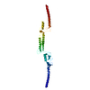
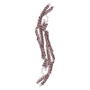
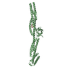
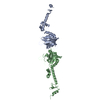
| Title | FCHO2 F-BAR domain | ||||||
 Components Components | FCH DOMAIN ONLY PROTEIN 2 | ||||||
 Keywords Keywords | LIPID BINDING PROTEIN / LIPID-BINDING PROTEIN / EFC DOMAIN /  VESICLE TRAFFICKING / MEMBRANE CURVATURE / ENDOCYTOSIS / EXOCYTOSIS / F-BAR DOMAIN / POLYMORPHISM / LIPID- BINDING PROTEIN / COILED-COIL VESICLE TRAFFICKING / MEMBRANE CURVATURE / ENDOCYTOSIS / EXOCYTOSIS / F-BAR DOMAIN / POLYMORPHISM / LIPID- BINDING PROTEIN / COILED-COIL | ||||||
| Function / homology |  Function and homology information Function and homology informationmembrane invagination / presynaptic endocytic zone membrane / clathrin coat assembly / clathrin-dependent endocytosis / membrane organization / clathrin-coated vesicle / phosphatidylserine binding / synaptic vesicle endocytosis / clathrin-coated pit / phosphatidylinositol-4,5-bisphosphate binding ...membrane invagination / presynaptic endocytic zone membrane / clathrin coat assembly / clathrin-dependent endocytosis / membrane organization / clathrin-coated vesicle / phosphatidylserine binding / synaptic vesicle endocytosis / clathrin-coated pit / phosphatidylinositol-4,5-bisphosphate binding / cytoskeletal protein binding / phosphatidylinositol binding / protein localization to plasma membrane / Cargo recognition for clathrin-mediated endocytosis / Clathrin-mediated endocytosis / cytoskeleton / identical protein binding / plasma membrane / cytosol / cytoplasmSimilarity search - Function | ||||||
| Biological species |  HOMO SAPIENS (human) HOMO SAPIENS (human) | ||||||
| Method | X-RAY DIFFRACTION / SYNCHROTRON / MIRAS / Resolution: 2.3 Å | ||||||
 Authors Authors | Henne, W.M. / McMahon, H.T. / Kent, H.M. / Evans, P.R. | ||||||
 Citation Citation | Journal: Structure / Year: 2007 Title: Structure and Analysis of Fcho2 F-Bar Domain: A Dimerizing and Membrane Recruitment Module that Effects Membrane Curvature. Authors: Henne, W.M. / Kent, H.M. / Ford, M.J.G. / Hedge, B.G. / Daumke, O. / Butler, P.J. / Mittal, R. / Langen, R. / Evans, P.R. / Mcmahon, H.T. | ||||||
| History |
|
- Structure visualization
| Structure viewer | Molecule: MolmilJmol/JSmol |
|---|
- Downloads & links
-Download
| PDBx/mmCIF format | 2v0o.cif.gz | 164.1 KB | Display | PDBx/mmCIF format |
|---|---|---|---|---|
| PDB format | pdb2v0o.ent.gz | 138.4 KB | Display | PDB format |
| PDBx/mmJSON format | 2v0o.json.gz | Tree view | PDBx/mmJSON format | |
| Others |  Other downloads Other downloads |
-Validation report
| Arichive directory | https://data.pdbj.org/pub/pdb/validation_reports/v0/2v0oftp://data.pdbj.org/pub/pdb/validation_reports/v0/2v0o | HTTPS FTP |
|---|
-Related structure data
| Similar structure data |
|---|


-Links
 PDBj
PDBj
- Assembly
| Deposited unit | 
| ||||||||||||
|---|---|---|---|---|---|---|---|---|---|---|---|---|---|
| 1 | 
| ||||||||||||
| 2 | 
| ||||||||||||
| Unit cell |
| ||||||||||||
| Noncrystallographic symmetry (NCS) | NCS oper:
|
-Components
| #1: Protein | Mass: 31527.719 Da / Num. of mol.: 3 / Fragment: F-BAR DOMAIN, RESIDUES 1-272 Source method: isolated from a genetically manipulated source Source: (gene. exp.) HOMO SAPIENS (human) / Plasmid: PGEX-6P1 / Production host:  ESCHERICHIA COLI (E. coli) / Strain (production host): BL21 / Variant (production host): PLYSS / References: UniProt: Q96CF5, UniProt: Q0JRZ9*PLUS ESCHERICHIA COLI (E. coli) / Strain (production host): BL21 / Variant (production host): PLYSS / References: UniProt: Q96CF5, UniProt: Q0JRZ9*PLUS#2: Chemical | Acetate  Mass: 59.044 Da / Num. of mol.: 3 / Source method: obtained synthetically / Formula: C2H3O2 Mass: 59.044 Da / Num. of mol.: 3 / Source method: obtained synthetically / Formula: C2H3O2#3: Water | ChemComp-HOH / | Water Mass: 18.015 Da / Num. of mol.: 180 / Source method: isolated from a natural source / Formula: H2O Mass: 18.015 Da / Num. of mol.: 180 / Source method: isolated from a natural source / Formula: H2OSequence details | FIRST 4 RESIDUES LGSP ARE A CLONING ARTEFACT FROM THE PLASMID | |
|---|
-Experimental details
-Experiment
| Experiment | Method: X-RAY DIFFRACTION / Number of used crystals: 1 |
|---|
- Sample preparation
| Crystal | Density Matthews: 3.7 Å3/Da / Density % sol: 66.97 % / Description: 2 SIMILAR HG DERIVATIVES |
|---|---|
| Crystal grow | pH: 9 / Details: 18% PEG4000, 300MM NA ACETATE, 100MM TRIS PH 9 |
-Data collection
| Diffraction | Mean temperature: 100 K |
|---|---|
| Diffraction source | Source: SYNCHROTRON / Site: ESRF  / Beamline: ID29 / Wavelength: 1 / Beamline: ID29 / Wavelength: 1 |
| Detector | Type: ADSC CCD / Detector: CCD / Date: Oct 7, 2006 |
| Radiation | Protocol: SINGLE WAVELENGTH / Monochromatic (M) / Laue (L): M / Scattering type: x-ray |
| Radiation wavelength | Wavelength: 1 Å / Relative weight: 1 |
| Reflection | Resolution: 2.3→45 Å / Num. obs: 61810 / % possible obs: 99.5 % / Observed criterion σ(I): -10 / Redundancy: 3.6 % / Rmerge(I) obs: 0.08 / Net I/σ(I): 10.2 |
| Reflection shell | Resolution: 2.3→2.42 Å / Redundancy: 3.7 % / Rmerge(I) obs: 0.94 / Mean I/σ(I) obs: 1.6 / % possible all: 99.2 |
- Processing
| Software |
| ||||||||||||||||||||||||||||||||||||||||||||||||||||||||||||||||||||||||||||||||||||||||||||||||||||||||||||||||||||||||||||||||||||||||||||||||||||||||||||||||||||||||||||||||||||||
|---|---|---|---|---|---|---|---|---|---|---|---|---|---|---|---|---|---|---|---|---|---|---|---|---|---|---|---|---|---|---|---|---|---|---|---|---|---|---|---|---|---|---|---|---|---|---|---|---|---|---|---|---|---|---|---|---|---|---|---|---|---|---|---|---|---|---|---|---|---|---|---|---|---|---|---|---|---|---|---|---|---|---|---|---|---|---|---|---|---|---|---|---|---|---|---|---|---|---|---|---|---|---|---|---|---|---|---|---|---|---|---|---|---|---|---|---|---|---|---|---|---|---|---|---|---|---|---|---|---|---|---|---|---|---|---|---|---|---|---|---|---|---|---|---|---|---|---|---|---|---|---|---|---|---|---|---|---|---|---|---|---|---|---|---|---|---|---|---|---|---|---|---|---|---|---|---|---|---|---|---|---|---|---|
| Refinement | Method to determine structure: MIRAS / Resolution: 2.3→119.52 Å / Cor.coef. Fo:Fc: 0.932 / Cor.coef. Fo:Fc free: 0.909 / SU B: 13.472 / SU ML: 0.293 / Cross valid method: THROUGHOUT / ESU R: 0.301 / ESU R Free: 0.252 / Stereochemistry target values: MAXIMUM LIKELIHOOD Details: HYDROGENS HAVE BEEN ADDED IN THE RIDING POSITIONS. THE ASYMMETRIC UNIT CONTAINS 1.5 DIMERS, ONE COMPRISING CHAINS A & B, AND THE OTHER CHAIN C WHICH FORMS A DIMER WITH ITS SYMMETRY MATE ...Details: HYDROGENS HAVE BEEN ADDED IN THE RIDING POSITIONS. THE ASYMMETRIC UNIT CONTAINS 1.5 DIMERS, ONE COMPRISING CHAINS A & B, AND THE OTHER CHAIN C WHICH FORMS A DIMER WITH ITS SYMMETRY MATE RELATED BY THE SYMMETRY OPERATOR (1-X,Y,1-Z).
| ||||||||||||||||||||||||||||||||||||||||||||||||||||||||||||||||||||||||||||||||||||||||||||||||||||||||||||||||||||||||||||||||||||||||||||||||||||||||||||||||||||||||||||||||||||||
| Solvent computation | Ion probe radii: 0.8 Å / Shrinkage radii: 0.8 Å / VDW probe radii: 1.2 Å / Solvent model: MASK | ||||||||||||||||||||||||||||||||||||||||||||||||||||||||||||||||||||||||||||||||||||||||||||||||||||||||||||||||||||||||||||||||||||||||||||||||||||||||||||||||||||||||||||||||||||||
| Displacement parameters | Biso mean: 65.93 Å2
| ||||||||||||||||||||||||||||||||||||||||||||||||||||||||||||||||||||||||||||||||||||||||||||||||||||||||||||||||||||||||||||||||||||||||||||||||||||||||||||||||||||||||||||||||||||||
| Refinement step | Cycle: LAST / Resolution: 2.3→119.52 Å
| ||||||||||||||||||||||||||||||||||||||||||||||||||||||||||||||||||||||||||||||||||||||||||||||||||||||||||||||||||||||||||||||||||||||||||||||||||||||||||||||||||||||||||||||||||||||
| Refine LS restraints |
|