 Movie
Movie Controller


+ Open data

- Basic information
| Entry | Database: PDB / ID: 2uxt | ||||||
|---|---|---|---|---|---|---|---|
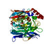
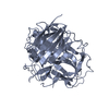
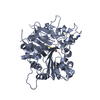
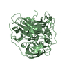
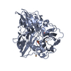
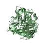
| Title | SufI Protein from Escherichia Coli | ||||||
 Components Components | PROTEIN SUFI | ||||||
 Keywords Keywords |  OXIDOREDUCTASE / SUFI / PERIPLASMIC / CUPREDOXIN-LIKE / FTS MUTANT SUPPRESSOR OXIDOREDUCTASE / SUFI / PERIPLASMIC / CUPREDOXIN-LIKE / FTS MUTANT SUPPRESSOR | ||||||
| Function / homology |  Function and homology information Function and homology informationFtsZ-dependent cytokinesis / response to ionizing radiation / cell division site / outer membrane-bounded periplasmic space / response to oxidative stress / oxidoreductase activity / copper ion binding / cell divisionSimilarity search - Function | ||||||
| Biological species |  ESCHERICHIA COLI (E. coli) ESCHERICHIA COLI (E. coli) | ||||||
| Method | X-RAY DIFFRACTION / SYNCHROTRON / MOLECULAR REPLACEMENT / Resolution: 1.9 Å | ||||||
 Authors Authors | Tarry, M.J. / Roversi, P. / Sargent, F. / Berks, B.C. / Lea, S.M. | ||||||
 Citation Citation | Journal: J.Mol.Biol. / Year: 2009 Title: The Escherichia Coli Cell Division Protein and Model Tat Substrate Sufi (Ftsp) Localizes to the Septal Ring and Has a Multicopper Oxidase-Like Structure. Authors: Tarry, M. / Arends, S.J. / Roversi, P. / Piette, E. / Sargent, F. / Berks, B.C. / Weiss, D.S. / Lea, S.M. | ||||||
| History |
| ||||||
| Remark 700 | SHEET THE SHEET STRUCTURE OF THIS MOLECULE IS BIFURCATED. IN ORDER TO REPRESENT THIS FEATURE IN ... SHEET THE SHEET STRUCTURE OF THIS MOLECULE IS BIFURCATED. IN ORDER TO REPRESENT THIS FEATURE IN THE SHEET RECORDS BELOW, TWO SHEETS ARE DEFINED. |
- Structure visualization
| Structure viewer | Molecule: MolmilJmol/JSmol |
|---|
- Downloads & links
-Download
| PDBx/mmCIF format | 2uxt.cif.gz | 182.3 KB | Display | PDBx/mmCIF format |
|---|---|---|---|---|
| PDB format | pdb2uxt.ent.gz | 143 KB | Display | PDB format |
| PDBx/mmJSON format | 2uxt.json.gz | Tree view | PDBx/mmJSON format | |
| Others |  Other downloads Other downloads |
-Validation report
| Arichive directory | https://data.pdbj.org/pub/pdb/validation_reports/ux/2uxtftp://data.pdbj.org/pub/pdb/validation_reports/ux/2uxt | HTTPS FTP |
|---|
-Related structure data
| Related structure data |  2uxvC  1pf3S C: citing same article ( S: Starting model for refinement |
|---|---|
| Similar structure data |


-Links
 PDBj
PDBj

- Assembly
| Deposited unit | 
| ||||||||
|---|---|---|---|---|---|---|---|---|---|
| 1 | 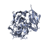
| ||||||||
| 2 | 
| ||||||||
| Unit cell |
| ||||||||
| Noncrystallographic symmetry (NCS) | NCS oper: (Code: given Matrix: (0.311, 0.791, 0.527), Vector : |
-Components
| #1: Protein | Mass: 50244.762 Da / Num. of mol.: 2 Source method: isolated from a genetically manipulated source Source: (gene. exp.) ESCHERICHIA COLI (E. coli) / Plasmid: P60-SUFI / Production host: ESCHERICHIA COLI (E. coli) / Strain (production host): M15 / Variant (production host): PREP4 / References: UniProt: P26648#2: Water | ChemComp-HOH / | Water Mass: 18.015 Da / Num. of mol.: 406 / Source method: isolated from a natural source / Formula: H2O Mass: 18.015 Da / Num. of mol.: 406 / Source method: isolated from a natural source / Formula: H2OSequence details | 6-HIS TAGGED AT THE CTERMINUS WITH ARG-SER LINKER | |
|---|
-Experimental details
-Experiment
| Experiment | Method: X-RAY DIFFRACTION / Number of used crystals: 1 |
|---|
- Sample preparation
| Crystal | Density Matthews: 1.88 Å3/Da / Density % sol: 34.6 % / Description: NONE |
|---|---|
| Crystal grow | pH: 8 / Details: 2.35 M NACL, 0.1 M IMIDAZOLE PH 8.25 |
-Data collection
| Diffraction | Mean temperature: 120 K |
|---|---|
| Diffraction source | Source: SYNCHROTRON / Site: ESRF  / Beamline: ID29 / Wavelength: 0.974 / Beamline: ID29 / Wavelength: 0.974 |
| Detector | Type: ADSC CCD / Detector: CCD / Date: May 20, 2006 |
| Radiation | Protocol: SINGLE WAVELENGTH / Monochromatic (M) / Laue (L): M / Scattering type: x-ray |
| Radiation wavelength | Wavelength: 0.974 Å / Relative weight: 1 |
| Reflection | Resolution: 1.9→77.4 Å / Num. obs: 58949 / % possible obs: 98.4 % / Observed criterion σ(I): 0 / Redundancy: 3.4 % / Biso Wilson estimate: 1.688 Å2 / Rmerge(I) obs: 0.08 / Net I/σ(I): 11.3 |
| Reflection shell | Resolution: 1.9→2 Å / Redundancy: 2.9 % / Rmerge(I) obs: 0.4 / Mean I/σ(I) obs: 2.2 / % possible all: 98 |
- Processing
| Software |
| |||||||||||||||||||||||||||||||||||||||||||||||||||||||||||||||||||||||||||||||||||||||||||||||
|---|---|---|---|---|---|---|---|---|---|---|---|---|---|---|---|---|---|---|---|---|---|---|---|---|---|---|---|---|---|---|---|---|---|---|---|---|---|---|---|---|---|---|---|---|---|---|---|---|---|---|---|---|---|---|---|---|---|---|---|---|---|---|---|---|---|---|---|---|---|---|---|---|---|---|---|---|---|---|---|---|---|---|---|---|---|---|---|---|---|---|---|---|---|---|---|---|
| Refinement | Method to determine structure: MOLECULAR REPLACEMENT Starting model: PDB ENTRY 1PF3 Resolution: 1.9→39.84 Å / Isotropic thermal model: TNT BCORREL / Cross valid method: THROUGHOUT / σ(F): 0 / Stereochemistry target values: TNT PROTGEO / Details: STRUCTURE REFINED IN BUSTER-TNT VERSION BETA 2.1.1
| |||||||||||||||||||||||||||||||||||||||||||||||||||||||||||||||||||||||||||||||||||||||||||||||
| Solvent computation | Solvent model: BABINET SCALING / Bsol: 80 Å2 / ksol: 0.412 e/Å3 | |||||||||||||||||||||||||||||||||||||||||||||||||||||||||||||||||||||||||||||||||||||||||||||||
| Refinement step | Cycle: LAST / Resolution: 1.9→39.84 Å
| |||||||||||||||||||||||||||||||||||||||||||||||||||||||||||||||||||||||||||||||||||||||||||||||
| Refine LS restraints |
|
